
Page 148 - Read Online
P. 148
Rao. Vessel Plus 2022;6:26 https://dx.doi.org/10.20517/2574-1209.2021.93 Page 17 of 43
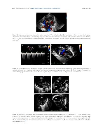
Figure 28. Suprasternal notch crab-view of the pulmonary veins (PV) draining into the left atrium (LA) are shown by color flow imaging.
The LA is in the center with four PVs showing color flow; the upper PVs show blue color while both lower PVs show red color. Each of
the PVs is also pulse-Doppler interrogated during any routine study and normal Doppler velocity recorded (not shown). Reproduced
from Ref. [21] .
Figure 29. (A) A high tricuspid regurgitant Doppler flow velocity is shown and is suggestive of elevated pulmonary artery pressure in a
neonate with the infra-diaphragmatic type of total anomalous pulmonary venous connection. (B) Color Doppler flow mapping
demonstrating right-to-left shunting across the atrial septum. Reproduced from Ref. [21] . RA: Right atrium; LA: left atrium.
Figure 30. Selected video frames of a 2-dimensional echocardiogram in parasternal long- (A) and short- (B, C) axis and apical four
chamber (D) views demonstrating a large right atrium (RA), right ventricle (RV), and main pulmonary artery (MPA) in an infant with
total anomalous pulmonary venous connection. Note that the enlarged RV encroaches onto the left ventricle compressing it posteriorly
(A, C) and to the left (D). The common pulmonary vein (CPV) is located behind (A, B) and superior to the left atrium (LA) (D).
Reproduced from Ref. [24] .