
Page 150 - Read Online
P. 150
Rao. Vessel Plus 2022;6:26 https://dx.doi.org/10.20517/2574-1209.2021.93 Page 19 of 43
Figure 31. Selected video frames from suprasternal notch views in a baby with the infra-diaphragmatic type of total anomalous
pulmonary venous connection showing pulmonary veins (PV) draining into a common pulmonary vein (CPV) which is located posterior
to the left atrium (LA); 2-dimensional (A) and color flow mapping (B) images are shown. Modified from Ref. [24] .
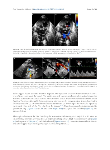
Figure 32. Selected video frames from suprasternal notch (A) and subcostal (B-D) views of an anomalous pulmonary venous trunk
(AT) draining the common pulmonary vein (CPV) in a baby with the infra-diaphragmatic type of total anomalous pulmonary venous
connection. A continuous wave Doppler recording of the obstructed anomalous trunk demonstrates increased velocity (D) suggesting
mild obstruction. Reproduced from Ref. [24] . LA: Left atrium.
Echo-Doppler studies provide a definitive diagnosis. The objective is to demonstrate the truncal anatomy,
type of truncus, status of the branch PAs (origin, size, and presence or absence of stenosis), intracardiac
anatomy, additional VSDs, aortic arch and other associated defects, and to evaluate the truncal valve and its
function. The echocardiographic features of truncus arteriosus are: (1) one great artery (truncus) originating
from the ventricles; (2) a VSD in the conal ventricular septum; (3) overriding of the ventricular septum by
the truncal valve; and (4) the PAs arise from the truncus [28-30] . These features are best demonstrated in
parasternal long [Figures 37A and 38] and short [Figure 37B] axis, apical four chamber [Figure 39], and
sub-costal views.
Thorough evaluation of the PAs, classifying the truncus into different types, namely, I, II or III based on
where the PAs arise and how they divide, is of paramount importance. High parasternal short axis [Figure
40] and suprasternal [Figure 41] and tilted subcostal [Figures 42 and 43] views with the use of both 2D echo
and color Doppler may help image the origin and branching of the PAs.