
Page 145 - Read Online
P. 145
Page 14 of 43 Rao. Vessel Plus 2022;6:26 https://dx.doi.org/10.20517/2574-1209.2021.93
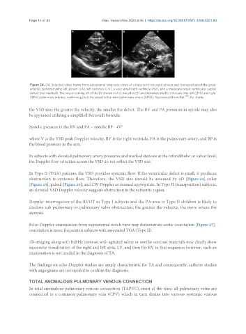
Figure 24. (A) Selected video frame from parasternal long-axis views of a baby with tricuspid atresia and transposition of the great
arteries demonstrating left atrium (LA), left ventricle (LV), a very small right ventricle (RV), and a moderate-sized ventricular septal
defect (not marked). The vessel coming off of the LV shown in A is traced in (B) and demonstrated to bifurcate into left (LPA) and right
(RPA) pulmonary arteries, confirming that this vessel is the main pulmonary artery (MPA). Reproduced from Ref. [19] . Ao: Aorta.
the VSD size; the greater the velocity, the smaller the defect. The RV and PA pressures in systole may also
be appraised utilizing a simplified Bernoulli formula:
Systolic pressure in the RV and PA = systolic BP - 4V 2
where V is the VSD peak Doppler velocity, RV is the right ventricle, PA is the pulmonary artery, and BP is
the blood pressure in the arm.
In subjects with elevated pulmonary artery pressures and marked stenosis at the infundibular or valvar level,
the Doppler flow velocities across the VSD do not reflect the VSD size.
In Type II (TGA) patients, the VSD provides systemic flow. If the ventricular defect is small, it produces
obstruction to systemic flow. Therefore, the VSD size should be assessed by 2D [Figure 24], color
[Figure 25], pulsed [Figure 26], and CW Doppler as deemed appropriate. In Type II (transposition) subjects,
an elevated VSD Doppler velocity suggests obstruction in the subaortic region.
Doppler interrogation of the RVOT in Type I subjects and the PA area in Type II children is likely to
disclose sub pulmonary or pulmonary valve obstruction; the greater the velocity, the more severe the
stenosis.
Echo-Doppler examination from suprasternal notch view may demonstrate aortic coarctation [Figure 27];
coarctation is more frequent in subjects with associated TGA (Type II).
2D imaging along with bubble contrast with agitated saline or similar contrast materials may clearly show
successive visualization of the right and left atria, LV, and then the RV in that sequence; however, such an
examination is not needed in the diagnosis of TA.
The findings on echo-Doppler studies are amply characteristic for TA and consequently, catheter studies
with angiograms are not needed to confirm the diagnosis.
TOTAL ANOMALOUS PULMONARY VENOUS CONNECTION
In total anomalous pulmonary venous connection (TAPVC), most of the time, all pulmonary veins are
connected to a common pulmonary vein (CPV) which in turn drains into various systemic venous