
Page 146 - Read Online
P. 146
Rao. Vessel Plus 2022;6:26 https://dx.doi.org/10.20517/2574-1209.2021.93 Page 15 of 43
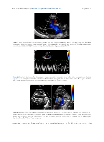
Figure 25. Selected video frame from parasternal long-axis view with color flow mapping of another patient with tricuspid atresia and
transposition of the great arteries demonstrates left atrium (LA), left ventricle (LV), a small right ventricle (RV), and a moderate-sized
ventricular septal defect (VSD). The vessel coming off the LV bifurcates. Reproduced from Ref. [19] .
Figure 26. Selected video frame of continuous wave Doppler across the ventricular septal defect of the same patient as shown in
[Figure 25]. Low velocity flow across the ventricular septal defect (VSD) suggests that the defect is non-obstructive. Reproduced from
Ref. [19] . Vmax: Maximum velocity; PG: peak gradient; RSVP: right ventricular systolic pressure.
Figure 27. Selected video frames from suprasternal notch views of the aortic (Ao) arch in 2D (A) and color flow (B) images of a
neonate with tricuspid atresia and transposition of the great arteries demonstrating coarctation of the aorta (CoA) and hypoplastic
transverse aortic arch (TAA). The association of CoA with tricuspid atresia plus transposition of the great arteries is well known.
Reproduced from Ref. [19] . DAo: Descending aorta.
structures. Less commonly, each pulmonary vein may directly connect to the RA, or the pulmonary veins