
Page 143 - Read Online
P. 143
Page 12 of 43 Rao. Vessel Plus 2022;6:26 https://dx.doi.org/10.20517/2574-1209.2021.93
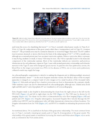
Figure 20. Selected video frames from subcostal long-axis views of the atrial septum, demonstrating a small color flow jet across the
patent foramen ovale (PFO; arrow in A), which was enlarged (arrow in B) after balloon atrial septostomy. The jet width is wider, and the
flow is laminar after balloon septostomy. Modified from Ref. [14] . LA: Left atrium; RA: right atrium.
and forms the source for classifying this lesion : (1) Type I, normally related great vessels; (2) Type II, d-
[15]
TGA; (3) Type III, malpositions of the great vessels other than d-transposition; and (4) Type IV, common
[15]
truncus . The proximal aorta may be normal in diameter or somewhat bigger than usual. The RV outflow
tract is normal, stenosed, or atretic; this forms the basis of the subgrouping of TA . A VSD is usually seen,
[15]
although an occasional patient may have an intact ventricular septum. When a ventricular defect is present,
it may be big, medium or small, or many VSDs may be seen. The VSD is typically located in the muscular
component of the ventricular septum. Most of the ventricular defects are restrictive and produce
obstruction in the sub pulmonary region in Type I cases with normal great artery relationship and subaortic
stenosis in the Type II cases with transposed great arteries. In patients who have pulmonary atresia, the
blood flow to the lungs is provided via a PDA; in the absence of PDA, one or more aorto-pulmonary
collateral vessels may supply the lungs.
An echocardiographic examination is valuable in making the diagnosis and in defining multiple structural
and hemodynamic issues [18-21] . In the most frequent muscular variety, the blocked tissue of the tricuspid
apparatus is imaged as a compact band of echo images at the usual location of the tricuspid valve
[Figure 21]. Subcostal and apical four-chamber projections are best to demonstrate the anatomic details.
Other anatomic types can also be demonstrated on echocardiography, although rare. Dilated atria (both
right and left) and LV and a hypoplastic RV are visualized on 2D echocardiography.
Echo-Doppler study is also helpful in demonstrating the shunt from the right atrium to the left via the
PFO/ASD [Figure 22] and left to right shunt via the VSD [Figure 23]. The VSD may be shown by 2D
imaging [Figure 23A]. The shunt via the defect is demonstrated by color flow imaging [Figure 23B] and
pulsed and CW [Figure 23C] Doppler interrogation. Increased Doppler flow velocity through the RV
outflow tract (RVOT) and the pulmonary valve will help demonstrate obstruction at these locations. The
Doppler information from the VSD [Figure 23C] and RVOT is valuable in estimating the pressures in the
PA.
As mentioned above, TA is classified on the basis of the great artery relationship. The most common forms
are: (1) Type I, normally related great vessels; and (2) Type II, d-transposition of the great vessels. The
relationship of the great vessels is established by tracing the vessels until bifurcation [Figure 24] or aortic
arch, as reviewed in the TGA section above.