
Page 138 - Read Online
P. 138
Rao. Vessel Plus 2022;6:26 https://dx.doi.org/10.20517/2574-1209.2021.93 Page 7 of 43
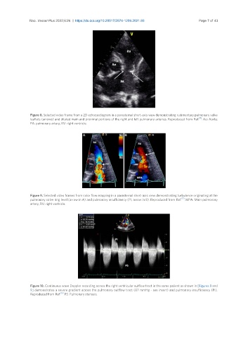
Figure 8. Selected video frame from a 2D echocardiogram in a parasternal short-axis view demonstrating rudimentary pulmonary valve
[5]
leaflets (arrows) and dilated main and proximal portions of the right and left pulmonary arteries. Reproduced from Ref. . Ao: Aorta;
PA: pulmonary artery; RV: right ventricle.
Figure 9. Selected video frames from color flow mapping in a parasternal short-axis view demonstrating turbulence originating at the
[5]
pulmonary valve ring level (arrow in A) and pulmonary insufficiency (PI; arrow in B). Reproduced from Ref. . MPA: Main pulmonary
artery; RV: right ventricle.
Figure 10. Continuous wave Doppler recording across the right ventricular outflow tract in the same patient as shown in [Figures 8 and
9] demonstrates a severe gradient across the pulmonary outflow tract (87 mmHg - see insert) and pulmonary insufficiency (PI).
[5]
Reproduced from Ref. . PS: Pulmonary stenosis.