
Page 134 - Read Online
P. 134
Rao. Vessel Plus 2022;6:26 https://dx.doi.org/10.20517/2574-1209.2021.93 Page 3 of 43
Figure 1. Selected video frame from a 2D echocardiogram in a parasternal long-axis view of a patient with tetralogy of Fallot
demonstrates a large ventricular septal defect (VSD) (thick arrow) and an over-riding aorta (Ao). The right ventricle (RV) is enlarged
[5]
and hypertrophied (RVH) (thin arrow). Reproduced from Ref. . LA: Left atrium; LV: left ventricle.
Figure 2. Color Doppler flow mapping of a similar echo frame as in [Figure 1], demonstrating right-to-left shunt (blue flow) across the
[5]
ventricular septal defect (VSD) (arrow). Other abbreviations are the same as in [Figure 1]. Reproduced from Ref. .
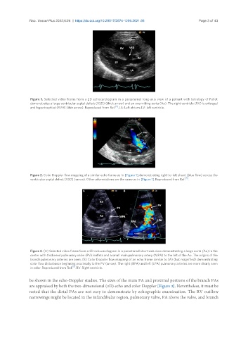
Figure 3. (A) Selected video frame from a 2D echocardiogram in a parasternal short-axis view demonstrating a large aorta (Ao) in the
center with thickened pulmonary valve (PV) leaflets and a small main pulmonary artery (MPA) to the left of the Ao. The origins of the
branch pulmonary arteries are seen. (B) Color Doppler flow mapping of an echo frame similar to (A) (but magnified) demonstrating
color flow disturbance beginning proximally to the PV (arrow). The right (RPA) and left (LPA) pulmonary arteries are more clearly seen
[5]
in color. Reproduced from Ref. . RV: Right ventricle.
be shown in the echo-Doppler studies. The sizes of the main PA and proximal portions of the branch PAs
are appraised by both the two-dimensional (2D) echo and color Doppler [Figure 3]. Nevertheless, it must be
noted that the distal PAs are not easy to demonstrate by echographic examination. The RV outflow
narrowings might be located in the infundibular region, pulmonary valve, PA above the valve, and branch