
Page 142 - Read Online
P. 142
Rao. Vessel Plus 2022;6:26 https://dx.doi.org/10.20517/2574-1209.2021.93 Page 11 of 43
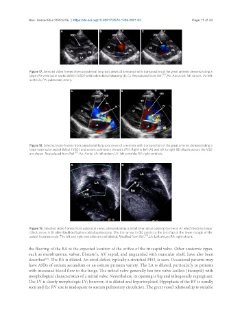
Figure 17. Selected video frames from parasternal long-axis views of a neonate with transposition of the great arteries demonstrating a
large (A) ventricular septal defect (VSD) with bidirectional shunting (B, C). Reproduced from Ref. [13] . Ao: Aorta; LA: left atrium; LV: left
ventricle; PA: pulmonary artery.
Figure 18. Selected video frames from parasternal long-axis views of a neonate with transposition of the great arteries demonstrating a
large ventricular septal defect (VSD) and severe pulmonary stenosis (PS). Right to left (A) and left to right (B) shunts across the VSD
are shown. Reproduced from Ref. [13] . Ao: Aorta; LA: left atrium; LV: left ventricle; RV: right ventricle.
Figure 19. Selected video frames from subcostal views, demonstrating a small inter-atrial opening (arrow in A) which became larger
(thick arrow in B) after Rashkind balloon atrial septostomy. The thin arrow in (B) points to the torn flap of the lower margin of the
[14]
patent foramen ovale. The left and right ventricles are not labeled. Modified from Ref. . LA: Left atrium; RA: right atrium.
the flooring of the RA at the expected location of the orifice of the tricuspid valve. Other anatomic types,
such as membranous, valvar, Ebstein’s, AV septal, and unguarded with muscular shelf, have also been
described . The RA is dilated. An atrial defect, typically a stretched PFO, is seen. Occasional patients may
[15]
have ASDs of ostium secundum or an ostium primum variety. The LA is dilated, particularly in patients
with increased blood flow to the lungs. The mitral valve generally has two valve leaflets (bicuspid) with
morphological characteristics of a mitral valve. Nevertheless, its opening is big and infrequently regurgitant.
The LV is clearly morphologic LV; however, it is dilated and hypertrophied. Hypoplasia of the RV is usually
seen and the RV size is inadequate to sustain pulmonary circulation. The great vessel relationship is variable