
Page 140 - Read Online
P. 140
Rao. Vessel Plus 2022;6:26 https://dx.doi.org/10.20517/2574-1209.2021.93 Page 9 of 43
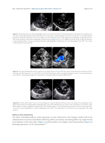
Figure 11. Parasternal long-axis echocardiographic views of two neonates, first, (A), with transposition of the great arteries and second,
(B), with normally related great arteries. Note that in (A), the posterior vessel arising from the left ventricle (LV) is coursing backward
(posteriorly) after its origin from the LV and is likely to be the pulmonary artery (PA), suggesting transposition of the great arteries.
This vessel should be traced and its bifurcation demonstrated (see Figure 12) to confirm that it is indeed the PA. In (B), the posterior
vessel arising from the LV courses somewhat anteriorly, indicating that it is likely to be the aorta. Reproduced from Ref. [13] . Ao: Aorta;
LA: left atrium; RV: right ventricle.
Figure 12. The vessel arising from the left ventricle of the infant shown in [Figure 11A] was traced, which showed bifurcation in 2D (A)
and color flow (B) images into the right (RPA) and left (LPA) pulmonary arteries suggesting that this vessel is the main pulmonary
artery (MPA), confirming the diagnosis of transposition of the great arteries. Reproduced from Ref. [13] .
Figure 13. Selected video frames from a 2D echocardiogram in long (A) and short (B) axis views of an infant with transposition of the
great arteries. Note the parallel position of the pulmonary artery (PA) and aorta (Ao) in A and on-end visualization of PA and Ao in (B).
These echoes are highly suggestive of transposition of the great arteries; in a normal baby, because of the normal crisscrossing of the
PA and Ao, such an appearance is unlikely. Reproduced from Ref. [13] . LA: Left atrium; LV: left ventricle; RA: right atrium; RV: right
ventricle.
Balloon atrial septostomy
The effect of Rashkind balloon atrial septostomy is easily evaluated by echo-Doppler studies both in the
catheterization laboratory immediately following balloon septostomy and during follow-up. Improvement
in 2D diameter of the atrial defect [Figure 19] and favorable color Doppler shunt characteristics [Figure 20]
following septostomy can be demonstrated [13,14] .