
Page 144 - Read Online
P. 144
Rao. Vessel Plus 2022;6:26 https://dx.doi.org/10.20517/2574-1209.2021.93 Page 13 of 43
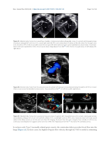
Figure 21. Selected video frames from apical four-chamber, 2-dimensional echocardiographic views of a patient with tricuspid atresia
showing an enlarged left ventricle (LV), a small right ventricle (RV), and a dense band of echoes at the site where the tricuspid valve
echo should be (ATV; thick arrow) with closed (A) and open (B) mitral valve during LV systole and diastole, respectively. A moderate-
sized ventricular septal defect (VSD; thin arrow) is shown. Reproduced from Ref. [19] . ATV: Atretic tricuspid valve; LA: left atrium; RA:
right atrium.
Figure 22. Selected video frame from the subcostal view of a patient with tricuspid atresia demonstrating the right to left (R to L) shunt
(thick arrow) across the interatrial communication. Reproduced from Ref. [19] . LA: Left atrium; RA: right atrium.
Figure 23. Selected video frames from parasternal long-axis views of a patient with tricuspid atresia with normally related great arteries
demonstrating enlarged left atrium (LA) and left ventricle (LV), a small right ventricle (RV), and a moderate-sized ventricular septal
defect (VSD; thick arrow) on 2D (A) and color flow (B) imaging. Turbulent flow (B) with a Doppler flow velocity of 2.91 m/s by
continuous wave Doppler (C) suggests some restriction of the VSD. Reproduced from Ref. [19] . Ao: Aorta; PA: pulmonary artery.
In subjects with Type I (normally related great vessels), the ventricular defect provides blood flow into the
lungs [Figure 23]. In these cases, the highest Doppler flow velocity through the VSD is useful in estimating