
Page 151 - Read Online
P. 151
Page 20 of 43 Rao. Vessel Plus 2022;6:26 https://dx.doi.org/10.20517/2574-1209.2021.93
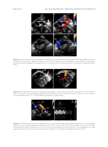
Figure 33. Selected video frames from suprasternal notch views in a neonate with the supra-diaphragmatic type of total anomalous
pulmonary venous connection showing a vertical vein (VV), left innominate vein (L Inn), and superior vena cava (SVC); 2-dimensional
(A, C) and color flow mapping (B, D) images are shown. Pulmonary veins (PV) draining into the VV are shown in B. Reproduced from
Ref. [24] .
Figure 34. Two-dimensional (A) and color flow mapping (B) images in a patient with a total anomalous pulmonary venous connection
to the coronary sinus (CS) demonstrating a dilated CS in 2D (A) and color (B). The pulmonary veins (PV) seem to connect to the CS
(B). Reproduced from Ref. [24] . LV: Left ventricle; RA: right atrium; RV: right ventricle.
Figure 35. (A) Selected video frame from suprasternal notch view in a neonate with the supra-diaphragmatic type of total anomalous
pulmonary venous connection showing a dilated vertical vein (VV), left innominate vein (L Inn), and superior vena cava (SVC). (B)
Continuous wave Doppler recordings from the VV show an increased Doppler flow velocity indicative of a mean gradient of 6 mmHg
suggestive of obstruction. CW: Continuous wave; PG: peak gradient; VTI: velocity time integral. Reproduced from Ref. [24] .