
Page 154 - Read Online
P. 154
Rao. Vessel Plus 2022;6:26 https://dx.doi.org/10.20517/2574-1209.2021.93 Page 23 of 43
Figure 42. Apical four chamber views [2D (A) and color Doppler (B)] with an anterior tilt of the transducer demonstrating the main
pulmonary artery (MPA) originating from the left side of the truncus (Tr). The MPA bifurcates into the right (RPA) and left (LPA)
pulmonary arteries. Reproduced from Ref. [28] .
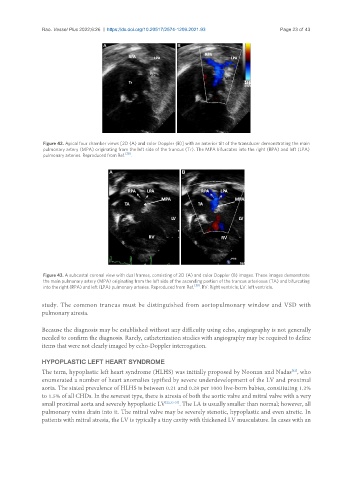
Figure 43. A subcostal coronal view with dual frames, consisting of 2D (A) and color Doppler (B) images. These images demonstrate
the main pulmonary artery (MPA) originating from the left side of the ascending portion of the truncus arteriosus (TA) and bifurcating
into the right (RPA) and left (LPA) pulmonary arteries. Reproduced from Ref. [30] . RV: Right ventricle; LV: left ventricle.
study. The common truncus must be distinguished from aortopulmonary window and VSD with
pulmonary atresia.
Because the diagnosis may be established without any difficulty using echo, angiography is not generally
needed to confirm the diagnosis. Rarely, catheterization studies with angiography may be required to define
items that were not clearly imaged by echo-Doppler interrogation.
HYPOPLASTIC LEFT HEART SYNDROME
The term, hypoplastic left heart syndrome (HLHS) was initially proposed by Noonan and Nadas , who
[32]
enumerated a number of heart anomalies typified by severe underdevelopment of the LV and proximal
aorta. The stated prevalence of HLHS is between 0.21 and 0.28 per 1000 live-born babies, constituting 1.2%
to 1.5% of all CHDs. In the severest type, there is atresia of both the aortic valve and mitral valve with a very
small proximal aorta and severely hypoplastic LV [22,33-37] . The LA is usually smaller than normal; however, all
pulmonary veins drain into it. The mitral valve may be severely stenotic, hypoplastic and even atretic. In
patients with mitral atresia, the LV is typically a tiny cavity with thickened LV musculature. In cases with an