
Page 157 - Read Online
P. 157
Page 26 of 43 Rao. Vessel Plus 2022;6:26 https://dx.doi.org/10.20517/2574-1209.2021.93
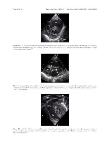
Figure 46. A selected frame from a 2D echocardiographic parasternal short-axis view in another neonate with hypoplastic left heart
syndrome demonstrates a small left ventricle (LV) and a large right ventricle (RV). A very small mitral valve (MV) (arrow) is also
shown. Reproduced from Ref. [21] .
Figure 47. A selected frame from a 2D echocardiographic parasternal long-axis view of a neonate with hypoplastic left heart syndrome
demonstrates a small left ventricle (LV), a markedly hypoplastic aorta (Ao) (arrow), and a large right ventricle (RV). Reproduced from
Ref. [21] . LA: Left atrium.
Figure 48. A selected video frame from a 2D echocardiographic apical four-chamber view in a neonate with hypoplastic left heart
syndrome, showing a hypoplastic left ventricle (LV) and markedly enlarged right ventricle (RV) and main pulmonary artery (MPA).
[21]
Reproduced from Ref. .