
Page 158 - Read Online
P. 158
Rao. Vessel Plus 2022;6:26 https://dx.doi.org/10.20517/2574-1209.2021.93 Page 27 of 43
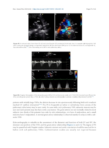
Figure 49. A selected video frame from 2D (A) and color Doppler (B) parasternal short-axis view in a neonate with hypoplastic left
heart syndrome demonstrating a somewhat constricted ductus arteriosus (DA) prior to the administration of prostaglandin E .
1
Reproduced from Ref. [21] . DAo: Descending aorta; MPA: main pulmonary artery.
Figure 50. Doppler interrogation of the descending aorta (DAo) (A) and transverse aortic arch (TAA) (B) showing forward flow in the
DAo and retrograde systolic flow in TAA; the latter finding indicates ductal-dependent systemic circulation. Reproduced from Ref. [21] .
patients with initially large VSDs, the defects decrease in size spontaneously following birth with resultant
marked LV outflow obstruction [40-42] . The PS is frequently at valvar or subvalvular level; atresia of the
pulmonary valve/artery may be seen rarely. In cases with a sub-pulmonary VSD, subaortic stenosis may be
seen and such patients may also have aortic coarctation. The great arteries are not normally related in most
subjects: two-thirds of the patients have side-by-side relationships, 25% have d-malposition, and a small
minority have l-malposition. A normal great artery relationship is observed mainly in subjects with a sub-
aortic VSD.
Echocardiography is valuable in the assessment of the diameter and function of both LV and RV, the
diameter and position of the VSD, and the great artery relationship [Figures 56 and 57]. The degree of PS
may be quantified with Doppler studies. Subaortic stenosis and aortic coarctation should be searched for in
babies with sub-pulmonary VSDs. Catheterization studies are usually not required because