
Page 159 - Read Online
P. 159
Page 28 of 43 Rao. Vessel Plus 2022;6:26 https://dx.doi.org/10.20517/2574-1209.2021.93
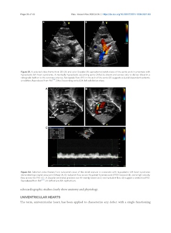
Figure 51. A selected video frame from 2D (A) and color Doppler (B) suprasternal notch views of the aortic arch in a neonate with
hypoplastic left heart syndrome. A markedly hypoplastic ascending aorta (AAo) is shown and serves only to deliver blood in a
retrograde fashion to the coronary arteries. Retrograde flow (RF) in the arch of the aorta (B) suggests a ductal-dependent systemic
circulation. Reproduced from Ref. [21] . DAo: Descending aorta; LSA: left subclavian artery.
Figure 52. Selected video frames from subcostal views of the atrial septum in a neonate with hypoplastic left heart syndrome
demonstrating a septal aneurysm (SAnu) (A, B), turbulent flow across the patent foramen ovale (PFO) (arrow in B), and a high-velocity
flow across the PFO (C). A Doppler-estimated gradient over 10 mmHg (insert in C) and turbulent flow (B) suggest a restrictive PFO.
Reproduced from Ref. [21] . LA: Left atrium; RA: right atrium.
echocardiographic studies clearly show anatomy and physiology.
UNIVENTRICULAR HEARTS
The term, univentricular heart, has been applied to characterize any defect with a single functioning