
Page 160 - Read Online
P. 160
Rao. Vessel Plus 2022;6:26 https://dx.doi.org/10.20517/2574-1209.2021.93 Page 29 of 43
Figure 53. Selected video frames from four-chamber two-dimensional echocardiographic views in five different neonates with varying
sizes of the left ventricle (LV) (A-E). Reproduced from Ref. [21] . LA: Left atrium; RA: right atrium; RV: right ventricle.
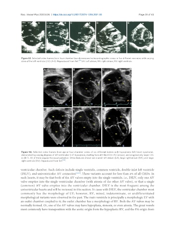
Figure 54. Selected video frames from apical four chamber views of six different babies with hypoplastic left heart syndrome,
demonstrating varying degrees of left ventricular (LV) hypoplasia, starting from slit-like LV in (A) (arrow), and progressively larger LVs
in (B-F). All of them require Norwood palliation. Other features shown are a small left atrium (LA), large right atrium (RA), and large
right ventricle (RV). Reproduced from Ref. [36] .
ventricular chamber. Such defects include single ventricle, common ventricle, double-inlet left ventricle
(DILV), and univentricular AV connection [22,39] . These variants account for less than 2% of all CHDs. In
such hearts, it may be that both of the AV valves empty into the single ventricle, i.e., DILV, only one AV
valve empties into the single ventricular chamber (with atresia of the other AV valve), or that a single
(common) AV valve empties into the ventricular chamber. DILV is the most frequent among the
univentricular hearts and will be reviewed in this section. In cases with DILV, the ventricular chamber most
commonly has the morphology of LV; however, RV, mixed, indeterminate, or undifferentiated
morphological variants were observed in the past. The main ventricle is principally a morphologic LV with
an outlet chamber coupled to it; the outlet chamber has a morphology of RV. Both the AV valves may be
normally formed. Or, one of the AV valves may have hypoplasia, stenosis, or even atresia. The great vessels
most commonly have transposition with the aortic origin from the hypoplastic RV, and the PA origin from