
Page 161 - Read Online
P. 161
Page 30 of 43 Rao. Vessel Plus 2022;6:26 https://dx.doi.org/10.20517/2574-1209.2021.93
Figure 55. Selected video frames from suprasternal notch views of four different babies (A-D) with hypoplastic left heart syndrome,
demonstrating varying degrees of ascending aortic (Ao) hypoplasia. Reproduced from Ref. [37] .
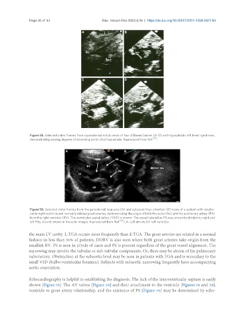
Figure 56. Selected video frames from the parasternal long-axis (A) and subcostal four chamber (B) views of a patient with double-
outlet right ventricle and normally related great arteries, demonstrating the origin of both the aorta (Ao) and the pulmonary artery (PA)
from the right ventricle (RV). The ventricular septal defect (VSD) is shown. The vessel labeled as PA was shown to divide into right and
left PAs, but not shown in the echo images. Reproduced from Ref. [39] . LA: Left atrium; LV: left ventricle.
the main LV cavity. L-TGA occurs more frequently than d-TGA. The great arteries are related in a normal
fashion in less than 30% of patients. DORV is also seen where both great arteries take origin from the
smallish RV. PS is seen in 2/3rds of cases and PS is present regardless of the great vessel alignment. The
narrowing may involve the valvular or sub-valvular components. Or, there may be atresia of the pulmonary
valve/artery. Obstruction at the subaortic level may be seen in patients with TGA and is secondary to the
small VSD (bulbo-ventricular foramen). Subjects with subaortic narrowing frequently have accompanying
aortic coarctation.
Echocardiography is helpful in establishing the diagnosis. The lack of the interventricular septum is easily
shown [Figure 58]. The AV valves [Figure 58] and their attachment to the ventricle [Figures 58 and 59],
ventricle to great artery relationship, and the existence of PS [Figure 59] may be determined by echo-