
Page 164 - Read Online
P. 164
Rao. Vessel Plus 2022;6:26 https://dx.doi.org/10.20517/2574-1209.2021.93 Page 33 of 43
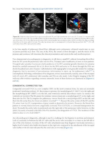
Figure 60. Selected video frames from supra-sternal notch (A) and high parasternal short axis (B, C) views in a neonate with
interrupted aortic arch. (A) The ascending (AAo) and descending (DAo) aortas are seen without any connection between them in this
and other views. (B, C) show the ductus arteriosus (DA) connecting the main pulmonary artery (MPA) with the DAo, thus providing
blood flow to the lower part of the body. The baby has already been on prostaglandin E infusion. Reproduced from Ref. [39] . LPA: Left
1
pulmonary artery; RPA: right pulmonary artery.
is the lone supply of pulmonary blood flow, although aorto-pulmonary collateral vessels may on rare
occasions provide such flow. The size of the PDA, the extent of flow through it, and the status of PA
pressure and resistance will determine the clinical presentation and course in the early newborn period.
Two-dimensional echocardiography is diagnostic; it will show a small RV without forwarding blood flow
from the RV across the pulmonary valve into the PAs. Tricuspid valve insufficiency is seen in most patients.
Tricuspid valve annulus Z scores are chiefly predictive of the RV’s potential growth; therefore, the annulus
should be carefully measured. RA to LA shunt via the PFO and aorta to PA shunt through the PDA are
shown by pulsed and color Doppler. Catheterization with angiography is not generally needed for making a
diagnosis, but is necessary if percutaneous intervention to open up the pulmonary valve [48,50] is
contemplated. Following confirmation of the diagnosis, several measurements, namely, sizes of the tricuspid
valve (Z score), RV, pulmonary valve annulus, and PAs are also made. Color Doppler imaging of the RV
wall may offer evidence regarding coronary sinusoids. Some of these features are illustrated in [Figures 61
and 62].
CONGENITAL CORRECTED TGA
Congenital corrected TGA is a rare complex CHD. In the most common form, the atria are normally
positioned (atrial situs solitus), AV discordance is present, the morphological LV (MLV) is to the right and
the morphological RV (MRV) is to the left, and ventriculo-arterial discordance exists. In essence, the
ventricles are inverted. The RA passes the systemic venous return into a morphological LV on the right, and
then into the PA. The LA empties the pulmonary venous blood into a morphological RV on the left, and
then into the aorta; thus, the blood circulation is normal [39,51-53] . Because the aorta comes off the RV and the
PA arises from the LV, transposition of great vessels is deemed to be present. However, the blood flow
pattern is normal, hence the term congenitally corrected TGA (CCTGA), or corrected transposition for
short. Normally the aortic valve is positioned onto the right side of the pulmonary valve; however, in this
defect, the aorta is located to the left of the PA; therefore, the designation of l-TGA. The incidence of this
defect is close to 0.5% of all CHDs .
[54]
An echocardiogram is diagnostic, although it may be a challenge for the beginner to perform and interpret.
Lack of continuity in-between the left AV valve and the aortic valve secondary to a conus on the left side is
one of the echo features. Scrutiny of the AV valve attachment may help diagnose ventricular inversion; a
higher level of mitral valve leaflet attachment on the right side compared to the attachment of the tricuspid
valve leaflet on the left side indicates that the ventricles are inverted [Figure 63].