
Page 167 - Read Online
P. 167
Page 36 of 43 Rao. Vessel Plus 2022;6:26 https://dx.doi.org/10.20517/2574-1209.2021.93
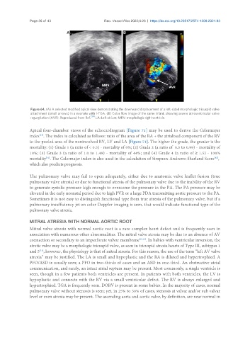
Figure 64. (A) A selected modified apical view demonstrating the downward displacement of a left-sided morphologic tricuspid valve
attachment (small arrows) in a neonate with l-TGA. (B) Color flow image of the same infant, showing severe atrioventricular valve
regurgitation (AVR). Reproduced from Ref. [39] . LA: Left atrium; MRV: morphologic right ventricle.
Apical four-chamber views of the echocardiogram [Figure 71] may be used to derive the Celermajer
index . The index is calculated as follows: ratio of the area of the RA + the atrialized component of the RV
[61]
to the pooled area of the noninvolved RV, LV and LA [Figure 71]. The higher the grade, the greater is the
mortality: (1) Grade 1 (a ratio of < 0.5) - mortality of 0%; (2) Grade 2 (a ratio of 0.5 to 0.99) - mortality of
10%; (3) Grade 3 (a ratio of 1.0 to 1.49) - mortality of 44%; and (4) Grade 4 (a ratio of ≥ 1.5) - 100%
mortality . The Celermajer index is also used in the calculation of Simpson-Andrews-Sharland Score ,
[62]
[61]
which also predicts prognosis.
The pulmonary valve may fail to open adequately, either due to anatomic valve leaflet fusion (true
pulmonary valve atresia) or due to functional atresia of the pulmonary valve due to the inability of the RV
to generate systolic pressure high enough to overcome the pressure in the PA. The PA pressure may be
elevated in the early neonatal period due to high PVR or a large PDA transmitting aortic pressure to the PA.
Sometimes it is not easy to distinguish functional type from true atresia of the pulmonary valve, but if a
pulmonary insufficiency jet on color Doppler imaging is seen, that would indicate functional type of the
pulmonary valve atresia.
MITRAL ATRESIA WITH NORMAL AORTIC ROOT
Mitral valve atresia with normal aortic root is a rare complex heart defect and is frequently seen in
association with numerous other abnormalities. The mitral valve atresia may be due to an absence of AV
connection or secondary to an imperforate valvar membrane [39,63] . In babies with ventricular inversion, the
atretic valve may be a morphologic tricuspid valve, as seen in tricuspid atresia hearts of Type III, subtypes 1
and 5 ; however, the physiology is that of mitral atresia. For this reason, the use of the term “left AV valve
[15]
atresia” may be justified. The LA is small and hypoplastic and the RA is dilated and hypertrophied. A
PFO/ASD is usually seen; a PFO in two-thirds of cases and an ASD in one-third. An obstructive atrial
communication, and rarely, an intact atrial septum may be present. Most commonly, a single ventricle is
seen, though in a few patients both ventricles are present. In patients with both ventricles, the LV is
hypoplastic and connects with the RV via a small ventricular defect. The RV is always enlarged and
hypertrophied. TGA is frequently seen. DORV is present in some babies. In the majority of cases, normal
pulmonary valve without stenosis is seen; yet, in 25% to 30% of cases, stenosis at valvar and/or sub valvar
level or even atresia may be present. The ascending aorta and aortic valve, by definition, are near normal in