
Page 171 - Read Online
P. 171
Page 40 of 43 Rao. Vessel Plus 2022;6:26 https://dx.doi.org/10.20517/2574-1209.2021.93
Figure 72. Selected video frames from modified apical four-chamber views of two infants (A, B) with mitral atresia, demonstrating
atretic mitral valves (AMV), indicated by thick arrows in (A) and (B). A small left atrium (LA) and left ventricle (LV), and a large right
ventricle (RV) are also seen. The thin arrow in (B) shows a restrictive patent foramen ovale (PFO). Reproduced from Ref. [39] .
Figure 73. Selected video frames from parasternal long-axis views of two infants (A, B) with mitral atresia, demonstrating atretic mitral
valves (AMV), indicated by thick arrows. In (B) a ventricular septal defect (VSD) is shown by a thin arrow. A small left ventricle (LV)
and a large right ventricle (RV) are also seen, particularly in (A). Reproduced from Ref. [39] .
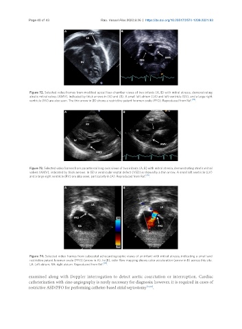
Figure 74. Selected video frames from subcostal echocardiographic views of an infant with mitral atresia, indicating a small and
restrictive patent foramen ovale (PFO) (arrow in A). In (B), color flow mapping shows color acceleration (arrow in B) across this site.
LA: Left atrium; RA: right atrium. Reproduced from Ref. [39] .
examined along with Doppler interrogation to detect aortic coarctation or interruption. Cardiac
catheterization with cine-angiography is rarely necessary for diagnosis; however, it is required in cases of
restrictive ASD/PFO for performing catheter-based atrial septostomy [14,63] .