
Page 168 - Read Online
P. 168
Rao. Vessel Plus 2022;6:26 https://dx.doi.org/10.20517/2574-1209.2021.93 Page 37 of 43
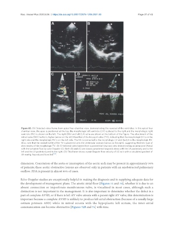
Figure 65. (A) Selected video frame from apical four-chamber view, demonstrating the reversal of the ventricles. In this apical four
chamber view, the apex is positioned at the top, the morphologic left ventricle (LV) is placed to the right and the morphologic right
ventricle (RV) is shown on the left. The right (RA) and left (LA) atria are shown at the bottom of the Figure. The attachment of the
mitral valve (MV) leaflet is higher (arrow on the left) than that of the tricuspid valve (TV), indicating that the morphologic LV is on the
right side and the morphologic RV is on the left side. The RA is connected to the morphologic LV and the LA to the morphologic RV.
Also, note that the medial leaflet of the TV is plastered onto the ventricular septum (arrow on the right), suggesting Ebstein’s type of
abnormality of the morphologic TV. (B, C) Selected video frames from a parasternal long-axis view demonstrating an aneurysm (Aneu)
with the turbulent flow by color Doppler (C). Both (B) and (C) are classic parasternal long-axis views with the LA posteriorly and to the
left and the LV posteriorly and to the right. (D) This frame shows a peak Doppler flow velocity of 3.5 m/s with a calculated gradient of
49 mmHg. Reproduced from Ref. [52] .
dimension. Coarctation of the aorta or interruption of the aortic arch may be present in approximately 30%
of patients; these aortic obstructive lesions are observed only in patients with an unobstructed pulmonary
outflow. PDA is present in almost 80% of cases.
Echo-Doppler studies are exceptionally helpful in making the diagnosis and in supplying adequate data for
the development of management plans. The atretic atrial floor [Figures 72 and 73], whether it is due to an
absent connection or imperforate membranous valve, is visualized in most cases, although such a
distinction is not important in the management. It is also important to determine whether the defect is a
part of complete AVSD, or if there is left AV valve atresia with a patent right AV valve; this determination is
important because a complete AVSD is unlikely to produce left atrial obstruction (because of a usually large
ostium primum ASD) while in mitral atresia with the hypoplastic left atrium, the inter-atrial
communication can become obstructive [Figures 72B and 74] with time.