
Page 169 - Read Online
P. 169
Page 38 of 43 Rao. Vessel Plus 2022;6:26 https://dx.doi.org/10.20517/2574-1209.2021.93
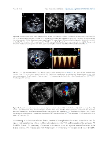
Figure 66. Selected video frames from right parasternal (A) and subcostal four-chamber (B) views of the morphological left ventricle
(MLV) showing an aneurysm (Anu) protruding into the pulmonary outflow tract, causing an obstruction. The Anu is located just below
the pulmonary valve (PV). A dilated main pulmonary artery (MPA) is also shown. (A) is a modified right parasternal view showing MLV
to the left, PV in the middle with an Anu below the PV. (B) is a modified subcostal four-chamber view showing MLV to the right and
Anu in the middle. (C) is a magnified view of (B), again illustrating the aneurysm and dilated MPA. Reproduced from Ref. [56] .
Figure 67. (A) Selected video frame from a subcostal four-chamber view (similar to Figure 66B) with color Doppler demonstrating
turbulent flow (TF) in the pulmonary outflow tract. (B) Continuous wave Doppler recording across the pulmonary outflow tract
[56]
demonstrates a peak Doppler velocity of approximately 5 m/s, suggesting significant obstruction. Reproduced from Ref. . MLV:
Morphological left ventricle.
Figure 68. Apical four-chamber views of an echocardiogram of a baby with a mild to moderate form of Ebstein’s anomaly. Panel (A)
shows a two-dimensional image demonstrating the insertion of the septal leaflet of the tricuspid valve (arrow), which is displaced
apically in comparison to the insertion of the mitral valve. The atrialized right ventricle (aRV) is shown. Panel (B) shows a color Doppler
image demonstrating moderate tricuspid valve regurgitation (TR). Reproduced from Ref. [58] . LA: Left atrium; LV: left ventricle; RA: right
atrium; RV: right ventricle.
The next step is to determine whether there is one ventricle (single ventricle) or two. In the latter case, the
type of ventricular looping (d loop vs. l loop), the diameter of the VSD, and the origins of the aorta and PA
should be defined. The pulmonary valve should be scrutinized to see if it is patent, stenosed or atretic and if
there is stenosis, CW Doppler may evaluate the degree of obstruction. Suprasternal notch views should be