Page 170 - Read Online
P. 170
Rao. Vessel Plus 2022;6:26  https://dx.doi.org/10.20517/2574-1209.2021.93       Page 39 of 43


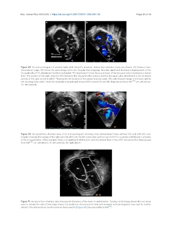
                Figure 69. An echocardiogram of another baby with Ebstein’s anomaly. Apical four-chamber views are shown. (A) Shows a two-
                dimensional image. (B) Shows the same image with color Doppler flow mapping. Note the significant downward displacement of the
                tricuspid valve (TV) attachment (solid arrow labeled “TV attachment”) from the true annulus of the tricuspid valve (marked by a dotted
                line). The portion of the right ventricle (RV) between the tricuspid valve annulus and the tricuspid valve attachment is the atrialized
                portion of the right ventricle (aRV). *Represents the location of the patent foramen ovale. The color Doppler image in B shows right to
                left shunting (blue color). Note the markedly enlarged right atrium (RA) in both (A) and (B). Reproduced from Ref. [60] . LA: Left atrium;
                LV: left ventricle.


                Figure 70. An apical five-chamber view of an echocardiogram showing a two-dimensional frame without (A) and with (B) color
                Doppler showing the bulging of the right ventricle (RV) into the left ventricular outflow tract (LVOT) in a patient with Ebstein’s anomaly
                of the tricuspid valve. In this example, there is no significant obstruction; note the laminar flow in the LVOT and aorta (Ao). Reproduced
                      [58]
                from Ref.  . LA: Left atrium; LV: left ventricle; RA: right atrium.


                Figure 71. An apical four-chamber view showing all chambers of the heart in end-diastole. Tracing on the image shows the two areas
                used to obtain the ratio (Celermajer index) for prediction of outcome in fetal and neonatal echocardiograms (see text for further
                details). The abbreviations are the same as those used in [Figure 68]. Reproduced from Ref. [58] .

