
Page 116 - Read Online
P. 116
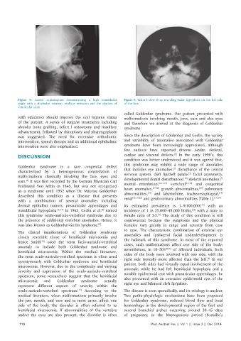
Figure 5:  Lateral cephalogram demonstrating a high mandibular   Figure 6: Water’s view X-ray revealing malar hypoplasia on the left side
          angle with a clockwise rotation, midface retrusion and the shadow of   of the face
          ventricular stunt
                                                              called Goldenhar syndrome. Our patient presented with
          with education should improve the oral hygiene status   malformations involving mouth, jaws, ears and also eyes
          of the patient. A series of surgical treatments including   and therefore we arrived at the diagnosis of Goldenhar
          alveolar bone grafting, Lefort I osteotomy and maxillary   syndrome.
          advancement, followed by rhinoplasty and pharyngoplasty
          was suggested. The need for extensive orthodontic   Since the description of Goldenhar and Gorlin, the variety
          intervention, speech therapy and an additional ophthalmic   and variability of anomalies associated with Goldenhar
          intervention were also emphasized.                  syndrome have been increasingly appreciated, although
                                                              few authors have reported diverse ocular, skeletal,
                                                                                       [7]
          DISCUSSION                                          cardiac and visceral defects.  In the early 1990’s, this
                                                              condition was better understood and it was agreed that,
                                                              this syndrome may exhibit a wide range of anomalies
                  Goldenhar syndrome is a rare congenital defect                       [7]
          characterized by a heterogeneous constellation of   that includes eye anomalies,  disturbance of the central
                                                                                              [7]
          malformations classically involving the face, eyes and   nervous system, cleft lip/cleft palate,  facial asymmetry,
                                                                                                              [7]
                                                                                            [12]
          ears.  It was first recorded by the German Physician     Carl   developmental dental disturbances,  skeletal anomalies,
              [4]
                                                                                     vertebral
                                                              mental retardation,
                                                                                             [14-16]
                                                                                                 and congenital
                                                                               [4,11,13]
          Ferdinand Von Arltin in 1845, but was not recognized   heart anomalies, [17-20]  growth abnormalities,  pulmonary
                                                                                                   [21]
          as a syndrome until 1952 when Dr. Maurice Goldenhar   abnormalities,  and labyrinthine, tracheoesophageal,
                                                                          [15]
                                                                                                             [15]
          described this condition as a disease that presents   renal [15,16,22]  and genitourinary abnormalities [Table 1]. [17,20]
          with a combination of several anomalies including
          dermal epibulbar tumors, preauricular appendages and   Its estimated prevalence is 1–9/100,000,  with an
                                                                                                    [13]
          mandibular hypoplasia. [3,5-7]  In 1963, Gorlin  et al.  named   incidence of 1 in 25,000–45,000 births,  with a male to
                                                                                                [28]
                                                   [8]
          this syndrome oculo-auriculo-vertebral syndrome due to   female ratio of 3:2.  The study of this condition is still
                                                                               [4]
          the presence of additional vertebral anomalies. Hence, it   controversial because the symptoms and the physical
          was also known as Goldenhar-Gorlin syndrome. [9]    features vary greatly in range and severity from case
                                                              to case. The characteristic combination of external ear
          The clinical manifestations of Goldenhar syndrome
          closely resemble those of hemifacial microsomia and   anomalies and ipsilateral facial underdevelopment is
          hence Smith  used the term facio-auriculo-vertebral   the hallmark of this syndrome. In most of the reported
                     [10]
          anomaly to include both Goldenhar syndrome and      cases, such malformations affect one side of the body;
                                                                                   [4,16]
          hemifacial microsomia. Within the medical literature,   nevertheless, in 10–50%   of affected individuals, both
          the term oculo-auriculo-vertebral spectrum is often used   sides of the body were involved with one side, with the
                                                                                                        [4]
          synonymously with Goldenhar syndrome and hemifacial   right side typically more affected than the left.  In our
          microsomia. However, due to the complexity and varying   patient, both sides had virtually equal involvement of the
          severity and expression of the oculo-auriculo-vertebral   anomaly; while he had left hemifacial hypoplasia and a
                                                              notable epidermoid cyst with preauricular appendages, he
          spectrum, some researchers suggest that the hemifacial   also presented with an extensive epidermoid cyst of the
          microsomia   and   Goldenhar   syndrome   actually  right eye and bilateral cleft lip/palate.
          represent different aspects of severity within the
          oculo-auriculo-vertebral spectrum.  According to the   The disease is seen sporadically, and its etiology is unclear.
                                        [11]
          medical literature, when malformations primarily involve   Two patho-physiologic mechanisms have been proposed
          the jaw, mouth, and ears and in most cases, affect one   for Goldenhar syndrome, reduced blood flow and focal
          side of the body, the disorder is often referred to as   hemorrhage in the developmental region of the first and
          hemifacial microsomia. If abnormalities of the vertebra   second branchial arches occurring around 30–45 days
          and/or the eyes are also present, the disorder is often   of pregnancy, in the blastogenesis period (Poswillo’s
           110                                                             Plast Aesthet Res || Vol 1 || Issue 3 || Dec 2014