
Page 115 - Read Online
P. 115
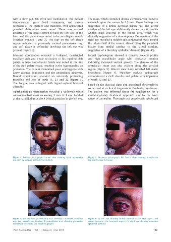
with a slow gait. On extra-oral examination, the patient   The mass, which contained dermal elements, was found to
          demonstrated gross facial asymmetry, and severe     encroach upon the cornea by 1.5 mm. These findings are
          retrusion of the midface and mandible. Well-demarcated   suggestive of a limbal dermoid [Figure 4a]. The lateral
          postcleft deformities were noted. There was marked   canthus of the left eye additionally showed a soft, mobile
          deviation of the nasal septum toward the left side of the   whitish mass growing in the bulbar area, which was
          face, and the patient was noted to be an obligate mouth   clinically suggestive of a dermolipoma. Examination of the
          breather [Figures 1 and 2]. The scar on the left cheek   right eye revealed a reddish sub-conjunctival mass around
          region indicated a previously excised preauricular tag,   the inferior half of the cornea, almost filling the palpebral
          and soft tissue is deformity involving the left ear was   fissure from medial canthus to the lateral canthus,
          present [Figure 2].                                 suggestive of a bleeding epibulbar dermoid [Figure 4b].
          Intraoral examination revealed a V-shaped, constricted   Lateral cephalogram showed a concave skeletal profile
          maxillary arch and a scar secondary to his repaired cleft   and high mandibular angle with clockwise rotation
          palate. A large nasoalveolar fistula was noted in the line   indicating increased vertical growth. The shadow of the
          of the cleft palate repair, resulting in the hypernasality on   ventricular shunt was also evident along the cervical
          pronation. The patient maintained poor oral hygiene with   region [Figure 5]. Water’s view X-ray revealed left malar
          heavy calculus deposition and the generalized gingivitis.   hypoplasia [Figure 6]. Maxillary occlusal radiograph
          Dental examination revealed an anteriorly protruding   demonstrated a cleft alveolus and palate with impaction
          mandible and loss of teeth 11, 21 and 22 [Figure 3].   of teeth 12 and 23.
          The tongue was enlarged with hypertrophied bilateral   Based on the classical signs and associated abnormalities
          adenoids.
                                                              we arrived at a clinical diagnosis of Goldenhar syndrome.
          Ophthalmologic examination revealed a yellowish white   The patient was informed about the requirement for a
          sub-conjunctival mass measuring 3 mm × 3 mm, located   multidisciplinary treatment approach due to the wide
          at the nasal limbus at the 9 O’clock position in the left eye.   range of anomalies. Thorough oral prophylaxis reinforced


          Figure 1:  Extroral photograph; frontal view showing facial asymmetry   Figure 2:  Extra-oral photograph; left lateral view showing preauricular
          and cleft lip surgery secondary deformity           tag and midface retrusion


                                                                          a
                     a


                     b                                                    b
          Figure 3:  Introral view. (a) Maxillary arch showing constricted maxillary   Figure 4:  (a) Left eye showing limbal dermoid in the nasal aspect and
          arch and nasoalveolar fistulae; (b) mandibular arch showing protrusive   dermo-lipoma in the temporal aspect; (b) right eye showing extensive
          mandibular anteriors and inflamed gingiva           epibulbar dermoid
          Plast Aesthet Res || Vol 1 || Issue 3 || Dec 2014                                                 109