
Page 118 - Read Online
P. 118
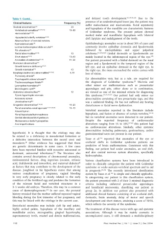
Table 1: Contd...                                   and delayed tooth development. [4,9,15,16,20]  Due to the
                                                              presence of an underdeveloped lower jaw, the patient may
           Clinical features                   Frequency (%)
                                                              suffer malocclusion and macrostomia. Facial asymmetry
           Skeletal abnormalities [20]              23        and hypoplasia of the mandible are characteristic features
            Vertebral abnormalities [4,14-16,24]   19–69      of Goldenhar syndrome. The present patient showed
            Hemivertebrae [15,24]
                                                              marked malar and mandibular hypoplasia with bilateral
            Hypoplastic/butterfl y vertebrae [15,24]           cleft lip/plate and malalignment of the teeth.
            Absence/fusion of cervical vertebra
            Kyphosis/scoliosis [15]                           Ophthalmologic anomalies occur in about 50% of cases and
            Lumbar lordosis/spina bifi da occulta [24]         commonly involve epibulbar dermoids and lipodermoids
            Rib alterations [24]                              followed by micropthalmia and upper palpebral
            Radial abnormalities [15,24]            13        coloboma. [16,20,24,25]  Limbal dermoids or lipodermoids are
                                                                                                             [25]
            Short stature [15,16]                  13–43      mainly located in the infratemporal region of the eye.
            Anomalies of extremities [15,16]       11–33      Our patient presented with a limbal dermoid on the nasal
           Hormonal abnormalities [21]                        region and a lipodermoid in the temporal region of the
            Growth hormone defi ciency [15,21]                 left eye, and an epibulbar dermoid in the right eye. In
            Growth retardation [15]                 43        the right eye, the mass encroached the entire cornea with
           Esophagic/pulmonary abnormalities [9,15,24]  6.70  loss of vision.
            Pulmonary atresia [9]
            Esophageal/duodenal atresia [24]                  Ear abnormalities vary, but as a rule, are required for
                                                                                                 [7]
            Tracheoesophageal fi stula [24]                    the diagnosis of Goldenhar syndrome.  Microtia and
            Laryngotracheomalacia [24]                        other minor ear malformations such as preauricular
            Bronchogenic cyst [24]                            appendages and pits, either alone or in combination,
           Abdominal abnormalities [20]             12        are viewed as one of the minimal criteria for diagnosing
            Pyloric hypertrophic stenosis                     this syndrome. [18,23,26,27]  Our patient had only minor ear
            Accessory spleen                                  abnormalities consisting of two preauricular tags, and this
            Umbilical hernia [24]                             was a unilateral finding. He has not suffered any hearing
           Urogenital abnormalities [7,16,20]      18–23      disturbances or facial nerve dysfunction.
            Renal anomalies-renal agenesis [7,15,16,22,24]  10–13
            Absent hymen                                      Vertebral anomalies reported in the literature include
                                                                                                            [14-16]
            Renal hypoplasia/hydronephrosis                   hypoplasia, and fusion or absence of certain vertebra,
            Genital alterations/short perineum                but no vertebral anomalies were detected in our patient.
            Maldescensus testis/hypospadias                   Despite the reported frequency of cardiovascular
            Anal imperforations                               alterations ranging from 5% to 58%, [17-20]  this patient had
                                                              no cardiovascular alterations. Similarly, other systemic
                                                              abnormalities including pulmonary, genitourinary, and/or
          hypothesis). It is thought that the etiology may also   gastrointestinal were not present in our patient.
          be related to a deficiency in mesodermal formation or
                                                                         [16]
          a defective interaction between the neural crest and   Tasse  et  al.  reported that anomalies of the eye or
          mesoderm.  Other evidence has suggested that there   orofacial clefts in Goldenhar syndrome patients are
                   [4]
          are genetic determinants in some cases. A few cases   predictive of brain malformations. Consistent with this
          have been reported families with recessive autosomal or   finding, our patient had ocular anomalies, an oral cleft,
          dominant, autosomal inheritance.  The literature also   and also central nervous system alteration, specifically
                                       [2]
          contains several descriptions of chromosomal anomalies,   hydrocephalus.
          environmental factors, drug ingestion (cocaine, retinoic   Various classification systems have been introduced in
          acid, thalidomide and temoxifen), and maternal diabetes    order to clinically categorize the patient with Goldenhar
                                                          [2]
          as factors that may contribute to the development of the   syndrome or oculo-auriculo-vertebral spectrum and to aid
          disease. Jongbloet in his hypothesis states that among   in prognosis. [16,25]  The recently introduced classification
          various complications of pregnancy, vaginal bleeding   system by Tasse et al.  is simple and clinically applicable.
                                                                                [16]
          in very early pregnancy is closely related to the early   In categorizing our patient in this classification system,
          condition of the fertilized egg and predates the formation   the patient presented with 2 of their 3 minimal diagnostic
          of the relevant facial and vertebral structures in a   criteria including unilateral preauricular appendages
          3–5 weeks old embryo. Therefore, this may be a common   and hemifacial microsomia, classifying our patient as
          cause of dysmorphogenesis.  In our case, the prenatal   group 2u. In addition our patient also presented with
                                  [18]
          history revealed that the mother suffered repeated vaginal   additional clinical features consisting of an orofacial cleft,
          bleeding during the first trimester of her pregnancy, and   dermoids, brain anomaly, delay of motor and speech
          this may be linked with the etiology in the current case.  development and short stature, attaining a score of 10/18,
                                                              which reflects the severity of the syndrome.
          Dentofacial anomalies may include cleft lip and palate,
          highly arched palate, hypoplasia of the maxillary and   The treatment of this disease varies with age and systemic
          mandibular arches, micrognathia, gingival hypertrophy,   associations. Although it may be mainly cosmetic in
          supernumerary teeth, enamel and dentin malformations,   uncomplicated cases, it still demands a multidisciplinary

           112                                                             Plast Aesthet Res || Vol 1 || Issue 3 || Dec 2014