
Page 121 - Read Online
P. 121
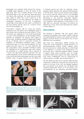
Radiographs were obtained which showed the absence   A Z-shaped incision was made for syndactyly release.
          of middle finger phalanges in both the hands. In the   Adequate thumb abduction was achieved intraoperatively.
          right hand, there was a variable absence of the third   In the left hand, central ray and transverse bony bridges
          metacarpal while in the left hand the third metacarpal   were excised, and cleft closure was done. The diverging
          was almost fully developed. Two small transverse bones   rays were held together employing a soft tissue sling
          were visible on either side of the central ray in the   functioning as the transverse metacarpal ligament.
          left hand [Figure 2]. No bony deficiency was visible on   Postoperatively mobilization and strengthening exercises
          radiographic examination of the feet. A hypoplastic   were instituted. Active abduction of the thumb on the
          second digit on the right foot was found to be devoid of   right hand was especially encouraged. The patient was
          the bony skeleton and the “great toe” consisted of first   followed for 3 years, with a good functional and cosmetic
          and second rays [Figure 3].                         outcome [Figure 5].

          Preanesthetic evaluation did not reveal any systemic
          involvement, but the patient had a Mallampatti grade II   DISCUSSION
          with normal neck movements and body weight of 22 kg.
          The patient was administered 5 mL syrup promethazine   The literature is abundant with case reports where
          as a part of the premedication. In the operating theater,   congenital abnormalities have always troubled surgeons
          induction of anesthesia was achieved with oxygen in   and anesthesiologists whenever such patients present for
          nitrous oxide, sevoflurane, propofol (2 mg/kg body weight),   surgical correction. However, in the present case, no such
          50  μg of fentanyl, and 2.5 vecuronium bromide for   difficulties were encountered.
          facilitation of endotracheal intubation using a cuffed   Split hand foot malformation (SHFM) involving the
          endotracheal tube of 5.5 mm internal diameter. The first   central rays of the extremities is usually inherited in an
          surgical procedure lasted for 2 h, with extubation and   autosomal-dominant fashion. Various causative genes
          the postoperative course uneventful. Diclofenac sodium   have been discovered, variable penetrance of which leads
          was administered for postoperative pain relief. Surgery   to the difference in severity of the manifestation. [7-9]  In
          was performed in a staged manner with an interval of   the present case, a thorough pedigree analysis revealed
          3 weeks. The right hand underwent operation first: the   a negative family history. Sporadic cases are usually
          central hypoplastic ray was excised, drill holes were made   encountered which show atypical cleft hands, which have
          in the second and fourth metacarpals, and the 2 were held   more severe manifestations and are characterized by
          together using a 22-gauge stainless steel wire [Figure 4].   variable absence of middle, ring and index fingers. [10]

                                                              The cleft divides the hand in two separate radial and ulnar
                                                              components. Sometimes a transverse bar of bone may be
                                                              present at the base of the cleft, but multiple fragments
                                                              were encountered in the present case. Syndactyly (most
                                                              commonly thumb-index finger, as in the present case)
                                                              and polydactyly are not uncommon associated findings.


          Figure 1:  Preoperative clinical photographs of the hand and feet; right
          hand showing syndactyly between the thumb and index finger; the
          right foot showing syndactyly between the third and fourth rays and a
          hypoplastic second toe; the left foot showing syndactyly between the   Figure 2:  Preoperative radiographs of the hands; note the accessory
          third and fourth toes                               transverse bony pieces in the cleft in the left hand (marked by arrows)


          Figure 3: Radiographs of both feet; the hypoplastic toe in the right foot
          has no bony scaffold                                Figure 4: Postoperative radiographs of both hands
          Plast Aesthet Res || Vol 1 || Issue 3 || Dec 2014                                                 115