
Page 125 - Read Online
P. 125
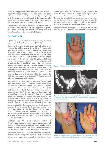
tumor in the hypothenar space and Guyon’s canal [Figure 3].   authors postulated that the lipoma originated from the
          The distal sensory branches of the ulnar nerve were firmly   nerve itself and termed it an “intra neural lipoma”. Our
          adherent to the tumor and were splayed by it. It appeared   case was similar in presentation. The lipoma was present
          as if the branches were embedded in the tumor capsule.   between the superficial and deep branches of the ulnar
          There was inadvertent injury to the ulnar digital nerve to   nerve. The superficial sensory branches were splayed by
          the little finger, which was repaired with 8–0 nylon.  the tumor and appeared to be embedded in the capsule.
                                                              Hence, an intraneural origin cannot be ruled out.
          Postoperative recovery was uneventful. The histopathological
          examination showed mature fat cells, suggestive of lipoma.   The possibility of nerve injury should always be discussed
          At 6-month follow-up, the patient was doing well with   with the patient preoperatively. Extreme caution should
          normal sensation on the ring and little fingers.

          DISCUSSION

          Lipoma in Guyon’s canal is rare, with only 13 cases
          reported, including the present case [Table 1].
                                            [9]
          Except for the case of an 8 years old,  all others were
          reported in adults, ranging from 36 to 74 years old,
          with a mean age of 52.2 years. There were 7 males and
          6 females. Nine of the 13 cases occurred on the right
          side. The tumor size varied from 1.5 cm × 1 cm (area)
          to 6.5 cm × 4 cm × 2.5 cm (volume), with this largest
          lesion seen in the present case. Six patients had only
          sensory involvement, 2 had only motor symptoms, and
          4 patients had a combined neuropathy. One patient
          had no neuropathy and was the only pediatric patient   Figure 1: Diffuse swelling in the hypothenar area
          in the series. This was attributed to unique anatomical
          and physiological differences for Guyon’s canal and the
          attending nerves.  MRI was taken in 7 cases. It gave
                         [9]
          accurate diagnosis in 5 patients, while, in 2 cases, the
          findings were suggestive of ganglion. [6,13]  Surgical removal
          alleviated symptoms in all patients.

          Shea and McClain have classified lesions of Guyon’s canal
          into three types: type I – proximal lesions having both
          sensory and motor involvement (30%), type II – lesions
          causing weakness of the intrinsic muscles (52%)
          and type III – distal lesions causing only sensory
          abnormalities (18%).  Recently Wu et al. have suggested
                           [14]
                                      [15]
          a classification into five types.  Type I is a mixed   a                      b
          motor and sensory neuropathy with the lesion at the   Figure 2: Magnetic resonance imaging finding of hyper-intense lesion in
          proximal end of Guyon’s canal. Type II is a pure sensory   T1 (a) with short T1 inversion recovery suppression (b) in hypothenar
                                                              space and Guyon’s canal
          neuropathy, with the lesion involving only the sensory
          branch. Type III is a pure motor neuropathy, with the
          lesion proximal to the branch supplying the hypothenar
          muscles. Type IV spares the hypothenar muscles with
          the lesion distal to the hypothenar muscle branch.
          Type V involves only the adductor pollicis and first
          dorsal interosseous muscles. The present case is type III
          according to Shea and McClain and type II according to
          the Wu classification.

          Ganglions  are  the   most   common    causes  of
          Guyon’s canal syndrome. Other causes include giant cell
          tumors, neurilemmomas, repetitive trauma, vascular lesions,
          anomalous muscles, carpal fractures and rheumatoid arthritis.
          Lipoma is a rare cause of nerve compression at this site.
          The cellular origin of lipoma in Guyon’s canal is debatable.   a               b
                         [10]
          Balakrishnan et al.  reported a case in which the branches   Figure 3:  Intra-operative pictures showing (a) dissection and (b)
          of the ulnar nerve were splayed by the tumor; these   enucleated specimen and underlying ulnar nerve and its branches

          Plast Aesthet Res || Vol 1 || Issue 3 || Dec 2014                                                 119