
Page 323 - Read Online
P. 323
Darbinyan et al. Neuroimmunol Neuroinflammation 2018;5:41 I http://dx.doi.org/10.20517/2347-8659.2018.33 Page 9 of 14
Dose 2.5 LD 50 , 1 h
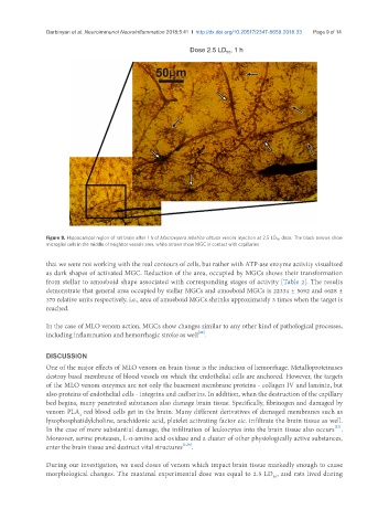
Figure 8. Hippocampal region of rat brain after 1 h of Macrovipera lebetina obtusa venom injection at 2.5 LD 50 dose. The black arrows show
microglial cells in the middle of neighbor vessels area, white arrows show MGC in contact with capillaries
that we were not working with the real contours of cells, but rather with ATP-ase enzyme activity visualized
as dark shapes of activated MGC. Reduction of the area, occupied by MGCs shows their transformation
from stellar to amoeboid shape associated with corresponding stages of activity [Table 2]. The results
demonstrate that general area occupied by stellar MGCs and amoeboid MGCs is 22334 ± 5092 and 6628 ±
370 relative units respectively, i.e., area of amoeboid MGCs shrinks approximately 3 times when the target is
reached.
In the case of MLO venom action, MGCs show changes similar to any other kind of pathological processes,
[26]
including inflammation and hemorrhagic stroke as well .
DISCUSSION
One of the major effects of MLO venom on brain tissue is the induction of hemorrhage. Metalloproteinases
destroy basal membrane of blood vessels on which the endothelial cells are anchored. However, the targets
of the MLO venom enzymes are not only the basement membrane proteins - collagen IV and laminin, but
also proteins of endothelial cells - integrins and cadherins. In addition, when the destruction of the capillary
bed begins, many penetrated substances also damage brain tissue. Specifically, fibrinogen and damaged by
venom PLA red blood cells get in the brain. Many different derivatives of damaged membranes such as
2
lysophosphatidylcholine, arachidonic acid, platelet activating factor etc. infiltrate the brain tissue as well.
[27]
In the case of more substantial damage, the infiltration of leukocytes into the brain tissue also occurs .
Moreover, serine proteases, L-α-amino acid oxidase and a cluster of other physiologically active substances,
enter the brain tissue and destruct vital structures [2,28] .
During our investigation, we used doses of venom which impact brain tissue markedly enough to cause
morphological changes. The maximal experimental dose was equal to 2.5 LD , and rats lived during
50