
Page 324 - Read Online
P. 324
Page 10 of 14 Darbinyan et al. Neuroimmunol Neuroinflammation 2018;5:41 I http://dx.doi.org/10.20517/2347-8659.2018.33
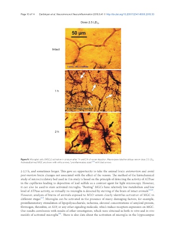
Dose 2.5 LD 50
Intact
1 h
2 h
Figure 9. Microglial cells (MGCs) activation in striatum after 1 h and 2 h of venom injection. Macrovipera lebetina obtusa venom dose 2.5 LD 50 .
Activated/primed MGC are shown with white arrows, “proinflammatory state” [39] with black arrows
2-2.5 h, and sometimes longer. This gave an opportunity to take the animal brain antemortem and avoid
post-mortem brain changes not associated with the effect of the venom. The method of the histochemical
study of microcirculatory bed used in this study is based on the principle of detecting the activity of ATPase
in the capillaries leading to deposition of lead sulfide as a contrast agent for light microscopy. However,
it can also be used to stain activated microglia. “Resting” MGCs have relatively low metabolism and low
level of ATPase activity, so virtually no microglia is detected by staining of the brain of intact animals [29,30] .
However, analysis of brains of animals exposed to MLO venom clearly identifies activation of MGC in
[31]
different stages . Microglia can be activated in the presence of many damaging factors, for example,
proinflammatory stimulation of lipopolysaccharide, ischemia, elevated concentrations of amyloid protein,
fibrinogen, thrombin, or ATP, or any other signaling molecule, which induce receptors expression on MGC.
Our results corroborate with results of other investigators, which were obtained in both in vitro and in vivo
models of activated microglia . There is also data about the activation of microglia in the hippocampus
[32]