
Page 819 - Read Online
P. 819
Page 8 of 23 Ancona et al. Mini-invasive Surg 2020;4:79 I http://dx.doi.org/10.20517/2574-1225.2020.80
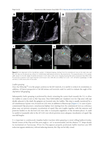
Figure 5. Axial alignment of the clip delivery system. A: biplane imaging, starting from the commissural view as the main view and
the LAX view as the derived one, allows for medial-lateral and anterior-posterior clip adjustments. The perpendicularity of the system
with respect to the MV plane should be achieved in both views; B-D: fusion imaging showing the same procedural step: RAO CRA
fluoroscopic projection with superimposed commissural view with (D) and without (C) color. LAA: left atrial appendage; LV: left
ventricle; LA: left atrium; MV: mitral valve
Leaflets grasping
TM
Once the Mitraclip is in the proper position in the left ventricle, it is useful to recheck its orientation; in
addition, 3D lateral perspective of the left atrium and ventricle could be useful to evaluate the angle of the
clip before full grasping.
Subsequently, leaflet grasping is performed by slowly retracting the system back towards the LA, to allow
the leaflets to come to rest on the Clip arms. Once both leaflets are visualized over the Clip arms with tips
ideally adjacent to the shaft, the grippers are lowered onto the leaflets. This step is usually monitored by a
2D simultaneous biplane view, focused on LAX view, in addition to fluoroscopy [Figure 8]. In cases of para-
commissural Clip placement, simultaneous biplane view starting from the commissural view as a reference
plane may not provide adequate visualization of equal Clip arm lengths together with the anterior and
posterior leaflets on the derived LAX view (due to inadequate angulation of the elevation plane). It could
be useful to transiently refer to the 2D LAX view which provides more adequate visualization of equal Clip
arm full lengths.
It is important to continuously visualize leaflet insertion while grasping to avoid rolling leaflets/chordae.
Partial closure of the Clip until the arms angle is ~60° is recommended, and this distinct “V” shape should
be maintained on fluoroscopy. When the Clip appears properly positioned, leaflet insertion and MR
reduction appear satisfactory without inducing stenosis, the Clip can be fully closed.