
Page 814 - Read Online
P. 814
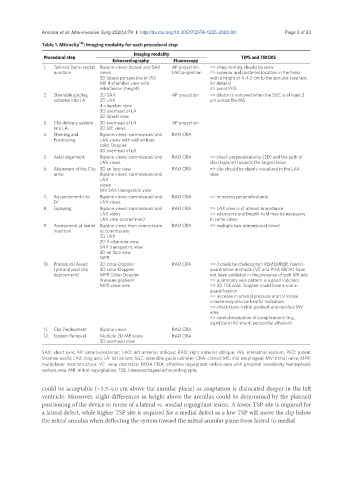
Ancona et al. Mini-invasive Surg 2020;4:79  I  http://dx.doi.org/10.20517/2574-1225.2020.80                                   Page 3 of 23

                            TM
               Table 1. Mitraclip : Imaging modality for each procedural step
                                              Imaging modality
                Procedural step                                                   TIPS and TRICKS
                                       Echocardiography     Fluoroscopy
                1.   Tailored Trans-septal  Biplane views: bicaval and SAX   AP projection  => sharp tenting should be seen
                   puncture        views                   LAO projection  => superior and posterior location in the fossa
                                   3D lateral perspective of IAS        with a height of 4-4.5 cm to the annulus (see text
                                   ME 4-chamber view with               for details)
                                   retroflexion (height)                => avoid PFO
                2.   Steerable guiding   2D SAX            AP projection  => dilator is removed when the SGC is at least 2
                   catheter into LA  2D LAX                             cm across the IAS
                                   4-chamber view
                                   3D overhead of LA
                                   3D lateral view
                3.   Clip delivery system   3D overhead of LA  AP projection
                   into LA         2D ME views
                4.   Steering and   Biplane views: commissural and   RAO CRA
                   Positioning     LAX views with and without
                                   color Doppler
                                   3D overhead of LA
                5.   Axial alignment  Biplane views: commissural and   RAO CRA  => check perpendicularity (3D) and the path of
                                   LAX views                            clip (biplane) towards the target lesion
                6.   Alignment of the Clip  3D en face view  RAO CRA    => clip should be clearly visualized in the LAX
                   arms            Biplane views: commissural and       view
                                   LAX
                                   views
                                   MV SAX transgastric view
                7.   Advancement into   Biplane views: commissural and   RAO CRA  => re-assess perpendicularity
                   LV              LAX views
                8.  Grasping       Biplane views: commissural and   RAO CRA  => LAX view is of utmost importance
                                   LAX views                            => adenosine and breath-hold may be necessary
                                   LAX view (sometimes)                 in some cases
                9.   Assessment of leaflet  Biplane views from commissure   RAO CRA  => multiple two-dimensional views!
                   Insertion       to commissure
                                   2D LAX
                                   2D 4-chamber view
                                   SAX transgastric view
                                   3D en face view
                                   MPR
                10.  Procedural Result   2D color-Doppler  RAO CRA      => it could be challenging!! REMEMBER: (semi)-
                   (pre and post clip   3D color-Doppler                quantitative methods (VC and PISA EROA) have
                   deployment)     MPR Color-Doppler                    not been validated in the presence of split MR jets
                                   Pressure gradient                    => pulmonary vein pattern is a good indicator
                                   MPR valve area                       => 3D TEE color Doppler could have a role in
                                                                        quantification
                                                                        => increase in arterial pressure and LV stroke
                                                                        volume may also be helpful indicators
                                                                        => check trans-mitral gradient and residual MV
                                                                        area
                                                                        => careful evaluation of complications (e.g.,
                                                                        significant IAS shunt, pericardial effusion)
                11.  Clip Deployment  Biplane views        RAO CRA
                12.  System Removal   Multiple 2D ME views  RAO CRA
                                   3D overhead view

               SAX: short axis; AP: antero-posterior; LAO: left anterior oblique; RAO: right anterior oblique; IAS: interatrial septum; PFO: patent
               foramen ovalis; LAX: long axis; LA: left atrium; SGC: steerable guide catheter; CRA: cranial; ME: mid esophageal; MV: mitral valve; MPR:
               multiplanar reconstruction; VC: vena contracta; EROA PISA: effective regurgitant orifice area with proximal isovelocity hemispheric
               surface area; MR: mitral regurgitation; TEE: transesophageal echocardiography


               could be acceptable (~3.5-4.0 cm above the annular plane) as coaptation is dislocated deeper in the left
               ventricle. Moreover, slight differences in height above the annulus could be determined by the planned
               positioning of the device in terms of a lateral vs. medial regurgitant lesion. A lower TSP site is required for
               a lateral defect, while higher TSP site is required for a medial defect as a low TSP will move the clip below
               the mitral annulus when deflecting the system toward the mitral annular plane from lateral to medial.