
Page 809 - Read Online
P. 809
Tredway et al. Mini-invasive Surg 2020;4:78 I http://dx.doi.org/10.20517/2574-1225.2020.77 Page 9 of 11
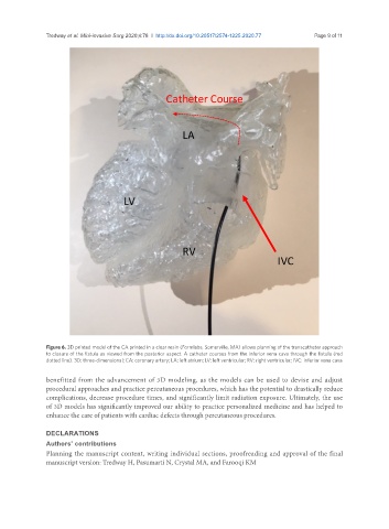
Figure 6. 3D printed model of the CA printed in a clear resin (Formlabs, Somerville, MA) allows planning of the transcatheter approach
to closure of the fistula as viewed from the posterior aspect. A catheter courses from the inferior vena cava through the fistula (red
dotted line). 3D: three-dimensional; CA: coronary artery; LA: left atrium; LV: left ventricular; RV: right ventricular; IVC: inferior vena cava
benefitted from the advancement of 3D modeling, as the models can be used to devise and adjust
procedural approaches and practice percutaneous procedures, which has the potential to drastically reduce
complications, decrease procedure times, and significantly limit radiation exposure. Ultimately, the use
of 3D models has significantly improved our ability to practice personalized medicine and has helped to
enhance the care of patients with cardiac defects through percutaneous procedures.
DECLARATIONS
Authors’ contributions
Planning the manuscript content, writing individual sections, proofreading and approval of the final
manuscript version: Tredway H, Pasumarti N, Crystal MA, and Farooqi KM