
Page 817 - Read Online
P. 817
Page 6 of 23 Ancona et al. Mini-invasive Surg 2020;4:79 I http://dx.doi.org/10.20517/2574-1225.2020.80
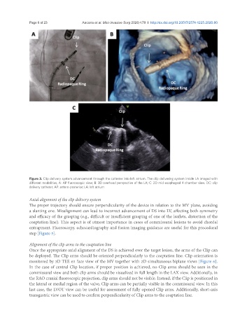
Figure 3. Clip delivery system advancement through the catheter into left atrium. The clip delivering system inside LA imaged with
different modalities, A: AP fluoroscopic view; B: 3D overhead perspective of the LA; C: 2D mid-esophageal 4 chamber view. DC: clip
delivery catheter; AP: antero-posterior; LA: left atrium
Axial alignment of the clip delivery system
The proper trajectory should ensure perpendicularity of the device in relation to the MV plane, avoiding
a slanting one. Misalignment can lead to incorrect advancement of DS into LV, affecting both symmetry
and efficacy of the grasping (e.g., difficult or insufficient grasping of one of the leaflets, distortion of the
coaptation line). This aspect is of utmost importance in cases of commissural lesions to avoid chordal
entrapment. Fluoroscopy, echocardiography and fusion imaging guidance are useful for this procedural
step [Figure 5].
Alignment of the clip arms to the coaptation line
Once the appropriate axial alignment of the DS is achieved over the target lesion, the arms of the Clip can
be deployed. The Clip arms should be oriented perpendicularly to the coaptation line. Clip orientation is
monitored by 3D TEE en face view of the MV together with 2D simultaneous biplane views [Figure 6].
In the case of central Clip location, if proper position is achieved, no Clip arms should be seen in the
commissural view and both clip arms should be visualized in full length in the LAX view. Additionally, in
the RAO cranial fluoroscopic projection, clip arms should not be visible. Instead, if the Clip is positioned in
the lateral or medial region of the valve, Clip arms can be partially visible in the commissural view. In this
last case, the LVOT view can be useful for assessment of fully opened Clip arms. Additionally, short-axis
transgastric view can be used to confirm perpendicularity of Clip arms to the coaptation line.