
Page 820 - Read Online
P. 820
Ancona et al. Mini-invasive Surg 2020;4:79 I http://dx.doi.org/10.20517/2574-1225.2020.80 Page 9 of 23
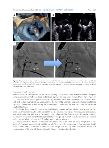
Figure 6. Alignment of the clip arms to the coaptation line. A: 3D MV en-face view guiding clip arms orientation with respect to the
coaptation line; B: biplane imaging: clip arms should be visible only in LAX view; C: RAO CRA fluoroscopic view: after adequate rotation
according to the echo guidance, clip arms are not visible anymore (right panel). LAX: long axis view; RAO CRA: right anterior oblique
cranial projection; MV: mitral valve
Assessment of leaflet insertion
The acquisition of a long loop is helpful as the grasping can be re-evaluated whenever needed. Adequate
leaflet insertion is verified when direct and indirect signs are simultaneously present. Direct signs [Figure 9]:
(1) The length of the leaflet captured inside the Clip should be ≥ 5 mm (some have reported at least 4 mm),
with both leaflets inserted into the atrial aspect of the closed Clip arms (the length of leaflet captured inside
the Clip is determined by subtracting the leaflet length outside the Clip from the corresponding leaflet
length at baseline);
(2) The leaflet draped over the closed arms should have a reduced mobility relative to the tips of the Clip
arms. Leaflets motility can be easily assessed by the 2D simultaneous biplane view, using the commissural
view as the main view and moving the elevation plane along the MV from the posterior-medial orifice to
the anterior-lateral one, closely to the edge of the Clip. The quality of insertion of the posterior and anterior
leaflets is usually best evaluated in LAX and 4-chamber view respectively;
(3) The occurrence of a double MV orifice: the 3D en face view of MV from LA or LV perspectives as well
as the 2D short axis transgastric view of MV are helpful to assess the new geometry of the valve and should
show a double-orifice valve with an adequate tissue bridge over the Clip arms. In addition, multiplanar
reconstruction of the same 3D dataset allows for further evaluation of the leaflets.