
Page 824 - Read Online
P. 824
Ancona et al. Mini-invasive Surg 2020;4:79 I http://dx.doi.org/10.20517/2574-1225.2020.80 Page 13 of 23
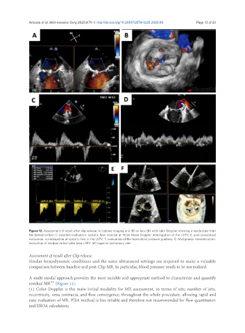
Figure 12. Assessment of result after clip release. A: biplane imaging and 3D en face (B) with color Doppler showing a residual jet from
the lateral orifice; C: baseline evaluation: systolic flow reversal at Pulse Wave Doppler interrogation of the LSPV; E: post-procedural
evaluation: normalization of systolic flow in the LSPV; F: evaluation of the transmitral pressure gradient; G: Multiplanar reconstruction:
evaluation of residual mitral valve area. LSPV: left superior pulmonary vein
Assessment of result after Clip release
Similar hemodynamic conditions and the same ultrasound settings are required to make a valuable
comparison between baseline and post-Clip MR. In particular, blood pressure needs to be normalized.
A multi-modal approach provides the most suitable and appropriate method to characterize and quantify
[11]
residual MR [Figure 12]:
(1) Color-Doppler is the main initial modality for MR assessment, in terms of site, number of jets,
eccentricity, vena contracta, and flow convergence, throughout the whole procedure, allowing rapid and
easy evaluation of MR. PISA method is less reliable and therefore not recommended for flow quantitation
and EROA calculation;