
Page 826 - Read Online
P. 826
Ancona et al. Mini-invasive Surg 2020;4:79 I http://dx.doi.org/10.20517/2574-1225.2020.80 Page 15 of 23
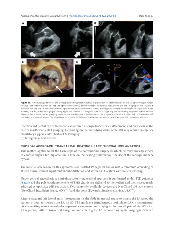
Figure 13. Procedural guidance of the transapical beating heart chordal implantation. A: identification of the LV apex through “finger
testing”: the interventionist pushes the apex (white arrow) and the imager checks its position on biplane imaging; B: the system is
directed towards the LA on simultaneous biplane LAX and commissural views, avoiding entrapment into subvalvular apparatus. After
entering the LA, echocardiographic imaging is switched to 3D surgical view (C), targeting the prolapsing segment (white arrow).
After confirmation of leaflet grasping and capture, the device is pulled out from the LV apex and tension is adjusted, until effective MR
reduction is shown with color Doppler interrogation (D). LV: left ventricular; LA: left atrium; LAX: long axis; MR: mitral regurgitation
insertion and partial clip detachment, also referred as single leaflet device attachment, and may occur in the
case of insufficient leaflet grasping. Depending on the underlying cause, acute MR may require emergency
circulatory support and/or bail-out MV surgery;
(5) Iatrogenic mitral stenosis.
CHORDAL APPROACH: TRANSAPICAL BEATING HEART CHORDAL IMPLANTATION
This method applies to all the basic steps of the conventional surgery in which delivery and adjustment
of chordal length after implantation is done on the beating heart without the use of the cardiopulmonary
bypass.
The most suitable lesion for this approach is an isolated P2 segment flail or with a minimum overriding of
at least 9 mm, without significant annular dilatation and severe LV dilatation with leaflets tethering.
Under general anaesthesia a mini-thoracotomy transapical approach is performed under TEE guidance
[Figure 13]: the polytetrafluorethylene (ePTFE) chords are delivered to the leaflets and then subsequently
adjusted to optimize MR reduction. Two currently available devices are NeoChord DS1000 system
[20]
(NeoChord, Inc., Eden Praire, MN) [17-19] and Harpoon (Edwards Lifescience, Irvine, USA) .
After a standard left lateral mini-thoracotomy in the fifth intercostal space to access the LV apex, the
system is directed towards the LA on 2D-TEE guidance (simultaneous multiplane LAX + commissural
views) avoiding native subvalvular apparatus entrapment and staying in the central part of the MV (A2-
P2 segments). After trans-mitral navigation and entering the LA, echocardiographic imaging is switched