Page 830 - Read Online
P. 830
Ancona et al. Mini-invasive Surg 2020;4:79  I  http://dx.doi.org/10.20517/2574-1225.2020.80                                   Page 19 of 23


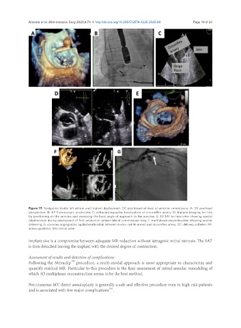
               Figure 17. Navigation inside left atrium and Implant deployment. DC positioned at level of anterior commissure: A: 3D overhead
               perspective; B: AP fluoroscopic projection; C: echocardiographic localization of circumflex artery; D: biplane imaging for fine
               tip positioning on the annulus and assessing the local angle of approach to the annulus; E: 3D MV en face view showing spatial
               relationships during deployment of first anchors in antero-lateral commissural area; F: multiplanar reconstruction showing anchor
               delivering; G: coronary angiography: spatial relationship between anchor (white arrow) and circumflex artery. DC: delivery catheter; AP:
               antero-posterior; MV: mitral valve

               implant size is a compromise between adequate MR reduction without iatrogenic mitral stenosis. The SAT
               is then detached leaving the implant with the desired degree of contraction.

               Assessment of results and detection of complications
                                    TM
               Following the Mitraclip  procedure, a multi-modal approach is most appropriate to characterize and
               quantify residual MR. Particular to this procedure is the final assessment of mitral annular remodeling of
               which 3D multiplanar reconstruction seems to be the best method.

               Percutaneous MV direct annuloplasty is generally a safe and effective procedure even in high-risk patients
                                                       [23]
               and is associated with few major complications .

