
Page 827 - Read Online
P. 827
Page 16 of 23 Ancona et al. Mini-invasive Surg 2020;4:79 I http://dx.doi.org/10.20517/2574-1225.2020.80
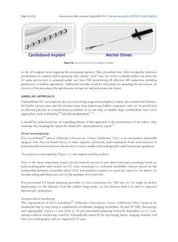
Figure 14. Components of the Cardioband System
to the 3D surgical view, targeting the prolapsing segment. This procedural step relies on specific technical
peculiarities to confirm leaflet grasping and capture. After that, the device is finally pulled out from the
LV apex and tension is adjusted under real time TEE monitoring till effective MR reduction, avoiding
asymmetry of leaflets apposition. Additional chordae could be implanted by repeating the procedure. At
the end of the procedure, the apical purse-strings are tied and access site closed.
ANNULAR APPROACH
Transcatheter MV annuloplasty devices, mimicking surgical annuloplasty, restore the normal ratio between
the leaflet surface area and the annular area, thus improving leaflets coaptation and can be performed
in selected patients as a stand-alone procedure or in one step or double steps combination with other
TM
approaches, such as Mitraclip /chordal implantation [21,22] .
It should be underlined that an appealing feature of this approach is the preservation of the native valve
[23]
anatomy, thus keeping the option for future MV interventions/re-repair .
Direct annuloplasty
The Cardioband device (Edwards Lifesciences, Irvine, California, USA) is an incomplete adjustable
TM
surgical-like Dacron band which is trans-septally delivered, and implanted from anterolateral to
posteromedial commissure on the posterior annulus under echocardiographic and fluoroscopic guidance.
The system is constituted by [Figure 14]: the implant and the anchors.
One of the most important aspect for procedural success is pre-interventional screening based on
echocardiography and mostly on CT scan, assessing (1) technical feasibility, mainly based on the
relationship between circumflex artery (CA) and posterior annulus to avoid the injury to the artery; (2)
annulus sizing and thickness; and (3) the anatomy of LA and IAS.
Pre-procedural CT based planning provides: (1) the coordinates for TSP site; (2) the angle of anchor
deployment; (3) the distance from the leaflets hinge point; (4) the distance from CA; and (5) expected
fluoroscopic projections.
Intraprocedural monitoring
The implantation of the Cardioband (Edwards Lifesciences, Irvine, California, USA) needs to be
TM
monitored step by step using a combination of different imaging modalities: 2D and 3D TEE, fluoroscopy
and angiography [Figure 15 and Table 2]. As pre-procedural planning is heavily dependent on CT scan,
intraprocedural monitoring could be tremendously eased by the upcoming fusion imaging between real
time echocardiography and pre-registered CT scan.