
Page 823 - Read Online
P. 823
Page 12 of 23 Ancona et al. Mini-invasive Surg 2020;4:79 I http://dx.doi.org/10.20517/2574-1225.2020.80
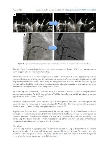
Figure 11. Clip release. Biplane imaging (A) and right anterior oblique fluoroscopic projections (B) showing clip release
The risk of mitral stenosis has to be evaluated by the assessment of diastolic TMPG via continuous-wave
(CW) Doppler after the placement of each Clip.
Planimetric assessment of the MV area provides an additive information. It should be preferably assessed
[8]
by using 3D imaging, which allows for multiplanar reconstruction . Alternatively, 2D planimetry could
be performed in the mid-diastole phase using the transgastric short-axis view. In both cases, the edges of
the MV leaflets should be clearly visible, allowing the inner edge of each orifice to be traced and the areas
added to calculate the total size of the newly formed orifices.
By combining both information, TMPG and MVA, it is possible to estimate the risk of iatrogenic mitral
2
stenosis more accurately. An MVA ≤ 1.5 cm and a TMPG ≥ 5 mmHg were considered criteria to indicate
significant MS in the EVEREST studies [9,10] .
Moreover, intraprocedural TMPG measured by TEE under general anesthesia conditions potentially
underestimates the hemodynamic impact of reduced MVA in daily life with exercise, which operators
[10]
should be aware of when deciding on implanting one or more clips .
Together with MVA and TMPG, the assessment of the final geometry of the MV should ensure: (1) each
clip is placed symmetrically on both leaflets and that the Clip is not biased towards one of them; and (2)
excessive distortion of the leaflets is avoided as it may lead to unbalanced traction and potentially cause
partial Clip detachment or leaflet rupture during follow-up. 3D en face view with atrial or ventricular
perspective is a fundamental imaging tool for this evaluation.
Clip release
Once the Clip position is appropriate and MR effectively reduced, the Clip is detached from the catheter
shaft usually under 2D imaging and fluoroscopic guidance [Figure 11]. A stable Clip position has to be
reconfirmed and the grade of residual MR should be reassessed by Color Doppler, as minor changes can
occur when the tension transferred via the DS disappears.