
Page 821 - Read Online
P. 821
Page 10 of 23 Ancona et al. Mini-invasive Surg 2020;4:79 I http://dx.doi.org/10.20517/2574-1225.2020.80
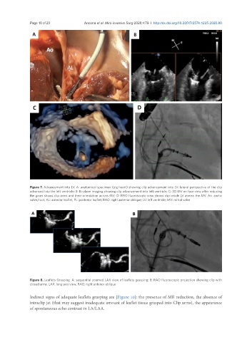
Figure 7. Advancement into LV. A: anatomical specimen (pig heart) showing clip advancement into LV: lateral perspective of the clip
advanced into the left ventricle; B: Bi-plane imaging showing clip advancement into left ventricle; C: 3D MV en face view after reducing
the gains shows clip arms and their orientation across MV; D: RAO fluoroscopic view shows clip inside LV across the MV. Ao: aortic
valve/root; AL: anterior leaflet, PL: posterior leaflet; RAO: right anterior oblique; LV: left ventricle; MV: mitral valve
Figure 8. Leaflets Grasping. A: sequential zoomed LAX view of leaflets grasping; B: RAO fluoroscopic projection showing clip with
closed arms. LAX: long axis view; RAO: right anterior oblique
Indirect signs of adequate leaflets grasping are [Figure 10]: the presence of MR reduction, the absence of
intraclip jet (that may suggest inadequate amount of leaflet tissue grasped into Clip arms), the appearance
of spontaneous echo contrast in LA/LAA.