
Page 818 - Read Online
P. 818
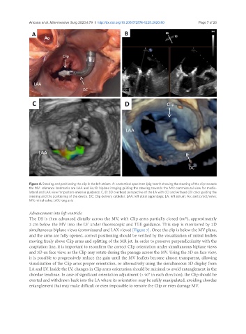
Ancona et al. Mini-invasive Surg 2020;4:79  I  http://dx.doi.org/10.20517/2574-1225.2020.80                                   Page 7 of 23


               Figure 4. Steering and positioning the clip in the left atrium. A: anatomical specimen (pig heart) showing the steering of the clip towards
               the MV: reference landmarks are LAA and Ao; B: biplane imaging guiding the steering towards the MV: commissural view for medio-
               lateral and LAX view for postero-anterior guidance; C, D: 3D overhead perspective of the LA with (C) and without (D) color guiding the
               steering and the positioning of the device. DC: Clip delivery catheter; LAA: left atrial appendage; LA: left atrium; Ao: aortic root/valve;
               MV: mitral valve; LAX: long axis

               Advancement into left ventricle
               The DS is then advanced distally across the MV, with Clip arms partially closed (60°), approximately
               2 cm below the MV into the LV under fluoroscopic and TEE guidance. This step is monitored by 2D
               simultaneous biplane views (commissural and LAX views) [Figure 7]. Once the clip is below the MV plane,
               and the arms are fully opened, correct positioning should be verified by the visualization of mitral leaflets
               moving freely above Clip arms and splitting of the MR jet. In order to preserve perpendicularity with the
               coaptation line, it is important to reconfirm the correct Clip orientation under simultaneous biplane views
               and 3D en face view, as the Clip may rotate during the passage across the MV. Using the 3D en face view,
               it is possible to progressively reduce the gain until the MV leaflets become almost transparent, allowing
               visualization of the Clip arms proper orientation, or alternatively using the simultaneous 3D display from
               LA and LV. Inside the LV, changes in Clip arms orientation should be minimal to avoid entanglement in the
               chordae tendinae. In case of significant orientation adjustment (> 90° in each direction), the Clip should be
               everted and withdrawn back into the LA where its orientation may be safely manipulated, avoiding chordae
               entanglement that may make difficult or even impossible to remove the Clip or even damage MV.