
Page 815 - Read Online
P. 815
Page 4 of 23 Ancona et al. Mini-invasive Surg 2020;4:79 I http://dx.doi.org/10.20517/2574-1225.2020.80
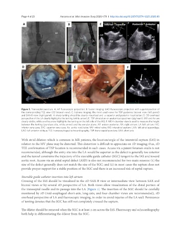
Figure 1. Transseptal puncture. A: AP fluoroscopic projection; B: fusion imaging: LAO fluoroscopic projection with superimposition of
the corresponding TEE view (2D bicaval view); C: biplane imaging: the most used views for TSP guidance: bicaval view (left panel)
and SAX-B view (right panel). A sharp tenting should be clearly visualized and a superior and posterior localization; D: 3D overhead
perspective of the LA clearly highlights the tenting (white arrow); E: TSP simulation on anatomical specimen (pig heart): MV and Ao are
clearly visible, while a withe arrow highlights the tenting on the left side of the IAS; F: ME 4-chamber view is used to measure the height
between the tenting (puncture site, white arrow) and the annular plane. AP: antero-posterior; RA: right atrium; LA: left atrium; IVC:
inferior vena cava; SVC: superior vena cava; Ao: aortic root/valve; MV: mitral valve; IAS: interatrial septum; LAA: left atrial appendage;
LAO: left anterior oblique; TEE: transesophageal echocardiography; TSP: trans-septal puncture; SAX: short axis
With atrial dilation which is common in MR patients, the location/angle of the interatrial septum (IAS) in
relation to the MV plane may be distorted. This distortion is difficult to appreciate on 2D imaging; thus, 3D
TEE confirmation of TSP location is recommended in such cases. Access via a patent foramen ovalis is not
recommended, although the entry site into the LA would be superior as the defect is generally too anterior
and the tunnel constrains the trajectory of the steerable guide catheter (SGC) tangent to the IAS and toward
aortic root. Access via an atrial septal defect (ASD) is also not recommended for two main reasons: (1) the
size of the defect generally does not match the size of the SGC; and (2) in most cases the septum does not
provide proper support for a stable position of the SGC and there is an increased risk of septal rupture.
Steerable guide catheter insertion into left atrium
Crossing of the IAS should be visualized in the 2D SAX-B view or intermediate view between SAX and
bicaval views or by several 3D perspective of LA. Both views allow visualization of the distal portion of
the transseptal needle and its passage into the LA [Figure 2]. The insertion of the SGC should be carefully
monitored by 2D (mid-esophageal short-axis, long-axis, and four-chamber views are recommended), 3D
overhead perspective of LA and fluoroscopic imaging, in order to avoid injuries of the LA wall. Persistence
of tenting denotes that the SGC has still not completely crossed the septum.
The dilator should be removed when the SGC is at least 2 cm across the IAS. Fluoroscopy and echocardiography
both help in differentiating the dilator from the SGC.