
Page 485 - Read Online
P. 485
Smer et al. Mini-invasive Surg 2020;4:52 I http://dx.doi.org/10.20517/2574-1225.2020.36 Page 7 of 15
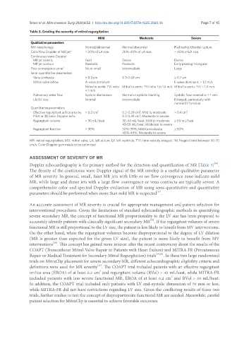
Table 3. Grading the severity of mitral regurgitation
Mild Moderate Severe
Qualitative parameters
MV morphology Normal/abnormal Normal/abnormal Flail leaflet/chordal rupture
Color flow Doppler of MR jet* < 20% of LA size 20%-40% of LA size > 40% of LA size
Continuous wave Doppler
MR jet density Faint Dense Dense
MR jet contour Parabolic Parabolic Early peaking-triangular
Flow convergence zone* No or small Intermediate Large
Semi-quantitative parameters
Vena contracta < 0.3 cm 0.3-0.69 cm ≥ 0.7 cm
Mitral valve inflow A-wave dominant E-wave dominant, > 1.2 m/s
Mitral to aortic TVI ratio Mitral to aortic TVI ratio 1 to 1.4 m/s Mitral to aortic TVI > 1.4 m/s
< 1 m/s
Pulmonary veins flow Systolic dominance Normal or systolic blunting Systolic flow reversal in > 1 vein
LA/LV size Normal Intermediate Enlarged, particularly with
normal LV function
Quantitative parameters
2
Effective regurgitant orifice area by < 0.2 cm 2 0.2-0.29 cm ; Mild to moderate ≥ 0.4 cm 2
2
PISA or 3D color Doppler echo 0.3-0.39 cm ; Moderate to severe
Regurgitant volume < 30 mL/beat 30-44 mL/beat; Mild to moderate ≥ 60 mL/beat
45-59 mL/beat; Moderate to severe
Regurgitant fraction < 30% 30%-39%; Mild to moderate ≥ 50%
40%-49%; Moderate to severe
MR: mitral regurgitation; MV: mitral valve; LA: left atrium; LV: left ventricle; TVI: time velocity integral. *At Nyquist limit between 50-70
cm/s. Color Doppler gain needs to be optimized
ASSESSMENT OF SEVERITY OF MR
Doppler echocardiography is the primary method for the detection and quantification of MR [Table 3] .
[16]
The density of the continuous wave Doppler signal of the MR envelop is a useful qualitative parameter
of MR severity. In general, small, faint MR jets with little or no flow convergence zone indicate mild
MR, while large and dense jets with a large flow convergence or vena contracta are typically severe. A
comprehensive color and spectral Doppler evaluation of MR using semi-quantitative and quantitative
[17]
parameters should be performed when more than mild MR is suspected .
An accurate assessment of MR severity is crucial for appropriate management and patient selection for
interventional procedures. Given the limitations of standard echocardiographic methods in quantifying
severe secondary MR, the concept of functional MR proportionality to the LV size has been proposed to
accurately identify patients with clinically significant secondary MR . If the regurgitant volumes of severe
[18]
functional MR is still proportional to the LV size, the patient is less likely to benefit from MV interventions.
On the other hand, when the regurgitant volumes become disproportional to the degree of LV dilation
(MR is greater than expected for the given LV size), the patient is more likely to benefit from MV
interventions . This concept has gained more interest after the recent controversy about the results of the
[18]
COAPT (Transcatheter Mitral-Valve Repair in Patients with Heart Failure) and MITRA-FR (Percutaneous
Repair or Medical Treatment for Secondary Mitral Regurgitation) trials [19,20] . In these two large randomized
trials on MitraClip placement for severe secondary MR, different echocardiographic eligibility criteria and
definitions were used for MR severity . The COAPT trial included patients with an effective regurgitant
[21]
2
orifice area (EROA) of at least 0.3 cm and regurgitant volume (RVol) > 45 mL/beat, while MITRA-FR
2
included patients with less severe functional MR, EROA of at least 0.2 cm and RVol > 30 mL/beat.
In addition, the COAPT trial included only patients with LV end-systolic dimension of 70 mm or less,
while MITRA-FR did not have restrictions regarding LV size. Given the conflicting results of these two
trials, further studies to test the concept of disproportionate functional MR are needed. Meanwhile, careful
patient selection for MitraClip is essential to achieve favorable outcomes.