
Page 483 - Read Online
P. 483
Smer et al. Mini-invasive Surg 2020;4:52 I http://dx.doi.org/10.20517/2574-1225.2020.36 Page 5 of 15
Table 2. Echocardiographic parameters for MitraClip feasibility
Favorable Unfavorable Contraindicated
Etiology of MR Myxomatous valve disease Severe annular dilation, > 50 mm or Rheumatic or endocarditis valve
EROA > 70.8 mm 2 disease
Location of MR Central, A2/P2 segments Peripheral, A1/P1 or A3/P3 segments Perforated mitral leaflets or clefts
Grasp zone
Calcification None Mild Moderate to severe
Length > 10 mm 7-10 mm < 7 mm
Mitral valve
Area > 4 cm 2 > 3.5 and < 4 cm 2 < 3.5 cm 2
Gradient < 4 mmHg > 4 and < 5 mmHg > 5 mmHg
Length of posterior leaflet > 10 mm 7-10 mm < 7 mm
Leaflet mobility Mobile Restricted motion Immobile
Primary MR Flail gap < 10 mm Flail gap > 10 mm
Flail width < 15 mm Flail width > 15 mm
Secondary MR Coaptation depth < 11 mm Coaptation depth > 11 mm
Coaptation length > 2 mm Coaptation length < 2 mm
EROA: effective regurgitation orifice area; MR: mitral regurgitation
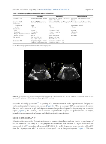
Figure 5. Two-dimensional transesophageal echocardiography demonstrates a flail MV (arrow) in five- and 2-chamber views. LV: left
ventricle; LA: left atrium; MV: mitral valve. Copyright with Aiman Smer
[14]
successful MitraClip placement . In primary MR, measurements of leaflet separation and flail gap and
width are important for procedural success [Figure 5]. While in secondary MR, measurements of annular
diameter and coaptation length and depth are essential to predict adequate leaflet grasping and successful
repair [Figure 6]. In addition, TEE is essential to guide both surgical and percutaneous MV repair,
immediately assess procedural success and identify potential complications.
3D ECHOCARDIOGRAPHY
3D echocardiography either from a transthoracic or transesophageal approach can provide superb images of
the MV apparatus. The ability of 3D imaging to visualize the MV from different 2D angles allows accurate
[15]
assessment of MR . A unique advantage of 3D TEE is the ability to provide an en face view of the MV
from the LA perspective, which is similar to the surgeon’s view in the operating room [Figure 7]. This view