
Page 486 - Read Online
P. 486
Page 8 of 15 Smer et al. Mini-invasive Surg 2020;4:52 I http://dx.doi.org/10.20517/2574-1225.2020.36
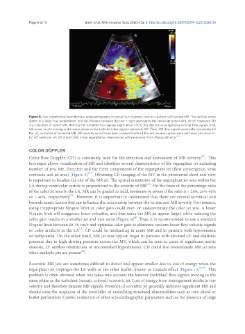
Figure 8. Two-dimensional transthoracic echocardiography in apical four-chamber view in a patient with severe MR. The vertical arrow
points to a large flow acceleration, and the distance between the two + signs represents the vena contracta width, which measures 1.13
cm, indicative of severe MR. Not only the turbulent flow signals (right arrow in LA) but also the accompanying laminar flow signals (red,
left arrow in LA) moving in the same phase as the turbulent flow signals represent MR. Thus, MR flow signals practically completely fill
the LA, indicative of torrential MR. MR severity would have been underestimated if the red laminar signals were not taken into account.
LV: left ventricle; LA: left atrium; MR: mitral regurgitation. Reproduced with permission from Manjunath et al. [4]
COLOR DOPPLER
[22]
Color flow Doppler (CD) is commonly used for the detection and assessment of MR severity . This
technique allows visualization of MR and identifies several characteristics of the regurgitant jet including
number of jets, site, direction and the three components of the regurgitant jet (flow convergence, vena
[1,4]
contracta and jet area) [Figure 8] . Obtaining CD imaging of the MV in the parasternal short-axis view
is important to localize the site of the MR jet. The spatial orientation of the regurgitant jet area within the
[23]
LA during ventricular systole is proportional to the severity of MR . On the basis of the percentage ratio
of the color jet area to the LA, MR can be graded as mild, moderate or severe if the ratio is < 20%, 20%-40%
or > 40%, respectively . However, it is important to understand that there are several technical and
[23]
hemodynamic factors that can influence the relationship between the jet size and MR severity. For instance,
using inappropriate Nyquist limit or color gain could over- or underestimate the color jet size. A lower
Nyquist limit will exaggerate lower velocities, and thus make the MR jet appear larger, while reducing the
[4]
color gain results in a smaller jet and vice versa [Figure 9] . Thus, it is recommended to use a standard
Nyquist limit between 50-70 cm/s and optimize color gain to eliminate random lower flow velocity signals
or color artifacts in the LA . CD could be misleading in acute MR and in patients with hypotension
[1]
or tachycardia. On the other hand, MR jet may appear larger in patients with elevated LV end-diastolic
pressure due to high driving pressure across the MV, which can be seen in cases of significant aortic
stenosis, LV outflow obstruction or uncontrolled hypertension. CD could also overestimate MR jet area
[24]
when multiple jets are present .
Eccentric MR jets are sometimes difficult to detect and appear smaller due to loss of energy when the
regurgitant jet impinges the LA walls or the other leaflet, known as Coanda effect [Figure 10] [4,25] . This
problem is often obviated when one takes into account the laminar (red/blue) flow signals moving in the
same phase as the turbulent (mosaic colored) eccentric jet. Loss of energy from impingement results in low
velocity and therefore laminar MR signals. Presence of eccentric jet generally indicates significant MR and
should raise the suspicion of the possibility of underlying structural abnormalities such as torn chord or
leaflet perforation. Careful evaluation of other echocardiographic parameters such as the presence of large