
Page 484 - Read Online
P. 484
Page 6 of 15 Smer et al. Mini-invasive Surg 2020;4:52 I http://dx.doi.org/10.20517/2574-1225.2020.36
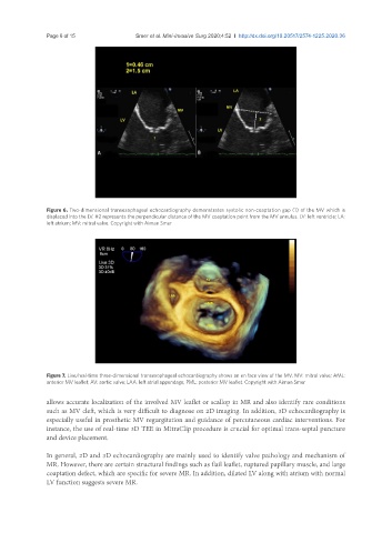
Figure 6. Two-dimensional transesophageal echocardiography demonstrates systolic non-coaptation gap (1) of the MV which is
displaced into the LV. #2 represents the perpendicular distance of the MV coaptation point from the MV annulus. LV: left ventricle; LA:
left atrium; MV: mitral valve. Copyright with Aiman Smer
Figure 7. Live/real-time three-dimensional transesophageal echocardiography shows an en face view of the MV. MV: mitral valve; AML:
anterior MV leaflet; AV: aortic valve; LAA: left atrial appendage; PML: posterior MV leaflet. Copyright with Aiman Smer
allows accurate localization of the involved MV leaflet or scallop in MR and also identify rare conditions
such as MV cleft, which is very difficult to diagnose on 2D imaging. In addition, 3D echocardiography is
especially useful in prosthetic MV regurgitation and guidance of percutaneous cardiac interventions. For
instance, the use of real-time 3D TEE in MitraClip procedure is crucial for optimal trans-septal puncture
and device placement.
In general, 2D and 3D echocardiography are mainly used to identify valve pathology and mechanism of
MR. However, there are certain structural findings such as flail leaflet, ruptured papillary muscle, and large
coaptation defect, which are specific for severe MR. In addition, dilated LV along with atrium with normal
LV function suggests severe MR.