
Page 480 - Read Online
P. 480
Page 2 of 15 Smer et al. Mini-invasive Surg 2020;4:52 I http://dx.doi.org/10.20517/2574-1225.2020.36
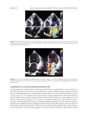
Figure 1. Two-dimensional transthoracic echocardiography demonstrates a myxomatous mitral valve and prolapse of posterior mitral
leaflet (arrow) with severe MR. RV: right ventricle; RA: right atrium; LV: left ventricle; LA: left atrium; MR: mitral regurgitation. Reproduced
with permission from Manjunath et al. [4]
Figure 2. Two-dimensional transthoracic echocardiography in apical four-chamber view shows a dilated LV with mitral valve coaptation
point displaced into the LV and severe MR. LV: left ventricle; LA: left atrium; MR: mitral regurgitation. Reproduced with permission from
[4]
Manjunath et al.
ASSESSMENT OF ETIOLOGY AND MECHANISM OF MR
Understanding the complex anatomy of the mitral valve (MV) is essential for accurate assessment of
MR. The MV apparatus consists of mitral annulus, MV leaflets, chordae tendineae, papillary muscles
and the underlying ventricular wall. Pathological abnormality of any one of these components can lead
[3,4]
to MR [Figure 1] . For instance, MR can occur due to primary (degenerative) MV disease affecting
the MV leaflets and/or chordae tendineae, while secondary MR occurs due to a pathological process of
[4,5]
the LV or left atrium (LA) [Figure 2] . In case of ventricular disease, due to either ischemic or non-
ischemic cardiomyopathy, MR occurs due to regional or global remodeling of the LV, which causes lateral
displacement of papillary muscles, resulting in annular dilation and leaflet tethering. However, there are
some differences in the mechanism of MR in these two types of cardiomyopathy. The main mechanism
for secondary MR in ischemic cardiomyopathy occurs due to inferior wall motion abnormalities, leading