
Page 481 - Read Online
P. 481
Smer et al. Mini-invasive Surg 2020;4:52 I http://dx.doi.org/10.20517/2574-1225.2020.36 Page 3 of 15
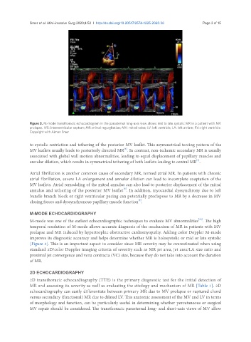
Figure 3. M-mode transthoracic echocardiogram in the parasternal long-axis view shows mid to late systolic MR in a patient with MV
prolapse. IVS: interventricular septum; MR: mitral regurgitation; MV: mitral valve; LV: left ventricle; LA: left atrium; RV: right ventricle.
Copyright with Aiman Smer
to systolic restriction and tethering of the posterior MV leaflet. This asymmetrical tenting pattern of the
[6]
MV leaflets usually leads to posteriorly directed MR . In contrast, non-ischemic secondary MR is usually
associated with global wall motion abnormalities, leading to equal displacement of papillary muscles and
[7]
annular dilation, which results in symmetrical tethering of both leaflets leading to central MR .
Atrial fibrillation is another common cause of secondary MR, termed atrial MR. In patients with chronic
atrial fibrillation, severe LA enlargement and annular dilation can lead to incomplete coaptation of the
MV leaflets. Atrial remodeling of the mitral annulus can also lead to posterior displacement of the mitral
[8]
annulus and tethering of the posterior MV leaflet . In addition, myocardial dyssynchrony due to left
bundle branch block or right ventricular pacing can potentially predispose to MR by a decrease in MV
[9]
closing forces and dyssynchronous papillary muscle function .
M-MODE ECHOCARDIOGRAPHY
M-mode was one of the earliest echocardiographic techniques to evaluate MV abnormalities . The high
[10]
temporal resolution of M-mode allows accurate diagnosis of the mechanism of MR in patients with MV
prolapse and MR induced by hypertrophic obstructive cardiomyopathy. Adding color Doppler M-mode
improves its diagnostic accuracy and helps determine whether MR is holosystolic or mid or late systolic
[Figure 3]. This is an important aspect to consider since MR severity may be overestimated when using
standard 2D/color Doppler imaging criteria of severity such as MR jet area, jet area/LA size ratio and
proximal jet convergence and vena contracta (VC) size, because they do not take into account the duration
of MR.
2D ECHOCARDIOGRAPHY
2D transthoracic echocardiography (TTE) is the primary diagnostic test for the initial detection of
MR and assessing its severity as well as evaluating the etiology and mechanism of MR [Table 1]. 2D
echocardiography can easily differentiate between primary MR due to MV prolapse or ruptured chord
versus secondary (functional) MR due to dilated LV. This anatomic assessment of the MV and LV in terms
of morphology and function, can be particularly useful in determining whether percutaneous or surgical
MV repair should be considered. The transthoracic parasternal long- and short-axis views of MV allow