
Page 489 - Read Online
P. 489
Smer et al. Mini-invasive Surg 2020;4:52 I http://dx.doi.org/10.20517/2574-1225.2020.36 Page 11 of 15
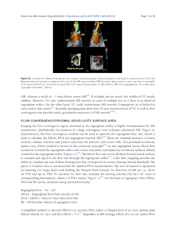
Figure 12. Live/real-time three-dimensional color Doppler transesophageal echocardiographic technique for assessment of VCA. The
three-dimensional dataset is cropped at the level of the MR vena contracta (MR jet origin) and cropped to view it en face (arrowhead).
2
VCA measured 0.43 cm , indicative of severe MR. VCA: vena contracta area; LV: left ventricle; MR: mitral regurgitation; AV: aortic valve.
Copyright with Navin C. Nanda
[22]
MR, whereas a width of > 7 mm defines severe MR . If multiple jets are noted, the widths of VC maybe
additive. However, VC may underestimate MR severity in cases of multiple jets or if there is an elliptical
regurgitant orifice. On the other hand, VC could overestimate MR severity if regurgitant jet is limited to
[29]
early, mid or late systole . Recently, emerging data show that 3D area measurements of VC as well as flow
convergence may provide useful quantitative assessment of MR severity [30,31] .
FLOW CONVERGENCE/PROXIMAL ISOVELOCITY SURFACE AREA
Imaging the flow convergence region proximal to the regurgitant orifice is highly recommended for MR
quantitation. Qualitatively, the presence of a large convergence zone indicates substantial MR [Figure 8].
Quantitatively, the flow convergence method can be used to quantify the regurgitant flow rate, which is
[32]
used to calculate the EROA, RVol and regurgitant fraction (RF) . These are essential measures of lesion
severity, volume overload and predict outcomes for patients with severe MR. The proximal isovelocity
[33]
surface area (PISA) method is based on the continuity principle . In any regurgitant lesion, blood flow
accelerates towards the regurgitant orifice and creates concentric hemispherical isovelocity surfaces (shells)
[34]
centered at the regurgitant orifice [Figure 13] . The blood flow rate across all these hemispherical surfaces
is constant and equal to the flow rate through the regurgitant orifice . Color flow mapping provides the
[33]
ability to visualize any one of these hemispheres that corresponds to certain aliasing velocity threshold. The
apical 4-chamber view is recommended for optimal PISA measurements. The area of interest is optimized
by lowering the image depth and shifting the Nyquist limit towards the direction of MR jet, i.e., down
on TTE and up on TEE. To calculate the flow rate, multiply the aliasing velocity (Va) by 2 πr (area of
2
[4]
corresponding hemisphere), where r is PISA radius [Figure 14] . On the basis of regurgitant flow, EROA,
RVol and RF can be calculated using standard formulas.
Regurgitant flow = Va × 2πr
2
EROA = Regurgitant flow/Peak velocity of MR
RVol = EROA × Velocity Time Interval of MR
RF = RVol/stroke volume of regurgitant valve
A simplified method to calculate EROA is to measure PISA radius at Nyquist limit of 40 cm/s, assume peak
2
[17]
2
MR jet velocity of 5 m/s, and then EROA = r /2 . Regardless of MR etiology, EROA of ≥ 0.4 cm and/or RVol